Biomarkers of immune response in mucosal lesions and their use with therapeutic vaccination

Inactive Publication Date: 2015-06-25
THE JOHN HOPKINS UNIV SCHOOL OF MEDICINE
View PDF0 Cites 1 Cited by
  • Summary
  • Abstract
  • Description
  • Claims
  • Application Information

AI Technical Summary

Benefits of technology

[0011]In accordance with an embodiment, the present invention provides a method for determining the effectiveness of an HPV vaccine regimen on a subject having a cervical intraepithelial neoplasia (CIN) comprising: a) obtaining at least one first tissue sample from the CIN of the subject; b) administering to the subject an effective amount of a first vaccine construct comprising HPV16 E7 antigen one or more times; c) administering to the subject an effective amount of a second vaccine construct comprising both E6 and E7 antigens from both HPV16 and HPV18; d) obtaining at least one or more second tissue samples from the CIN of the subject about 4 to about 24 weeks after step c); e)measuring the amount of CD8+ T cell infiltrates in the intraepithelial tissue of the first and second CIN samples; and f) determining that the HPV vaccine regimen is effective when the amount of CD8+ T cell infiltrates in the second tissue sample is increased when compared to the amount of CD8+ T cell infiltrates in the first tissue sample.
[0012]In accordance with an embodiment, the present invention provides a method for treating a subject having a cervical intraepithelial neoplasia (CIN) comprising: a) obtaining at least one first tissue sample from the CIN of the subject; b) administering to the subject an effective amount of a first vaccine construct comprising HPV16 E7 antigen one or more times; c) administering to the subject an effective amount of a second vaccine construct comprising both E6 and E7 antigens from both HPV16 and HPV18; d) obtaining at least one or more second tissue samples from the CIN of the subject about 4 to about 24 weeks after step c); e) measuring the amount of CD8+ T cell infiltrates in the intraepithelial tissue of the first and second CIN samples; and f) determining that the HPV treatment is effective when the amount of CD8+ T cell infiltrates in the second tissue sample is increased when compared to the amount of CD8+ T cell infiltrates in the first tissue sample.
[0013]In accordance with an embodiment, the present inve

Problems solved by technology

In the end, although much has been learned about the immunobiology of HPV-associated disease, the problem of translation of this knowledge into strategies to prevent and treat disease remains unresolved.
These observations suggested that, although CD8 T cells are likely to play a role in elimination of preinvasive, incipient HPV-associated neoplasia, access to lesional

Method used


Image

  • Biomarkers of immune response in mucosal lesions and their use with therapeutic vaccination
  • Biomarkers of immune response in mucosal lesions and their use with therapeutic vaccination
  • Biomarkers of immune response in mucosal lesions and their use with therapeutic vaccination

Examples


example 1

[0098]Heterologous prime-boost vaccination targeting HPV16 E6 / E7 is safe and tolerable in subjects with HPV16+ CIN2 / 3 lesions.

[0099]Healthy subjects with HPV 16-associated CIN2 / 3 underwent peripheral vaccination with a heterologous DNA prime-recombinant vaccinia vector-based boost vaccination regimen administered intramuscularly in the deltoid muscle before a standard therapeutic resection. The regimen included two priming vaccinations with a DNA vaccine expressing HPV16 E7 (DNAE7) at study weeks 0 and 4, followed by a recombinant vaccinia boost expressing HPV16 and HPV18 E6 and E7 (rVacE6E7; TA-HPV) at study week 8. At week 15, 7 weeks after the boost vaccination, subjects underwent a standard therapeutic resection of the cervical squamocolumnar junction. A total of 12 patients in three treatment arms were evaluated (Table 1).

[0100]Demographics are presented in Table 2. All subjects received 3 mg of DNAE7 at entry and at week 4. At week 8, patients received one of three rVacE6E7 do...

example 2

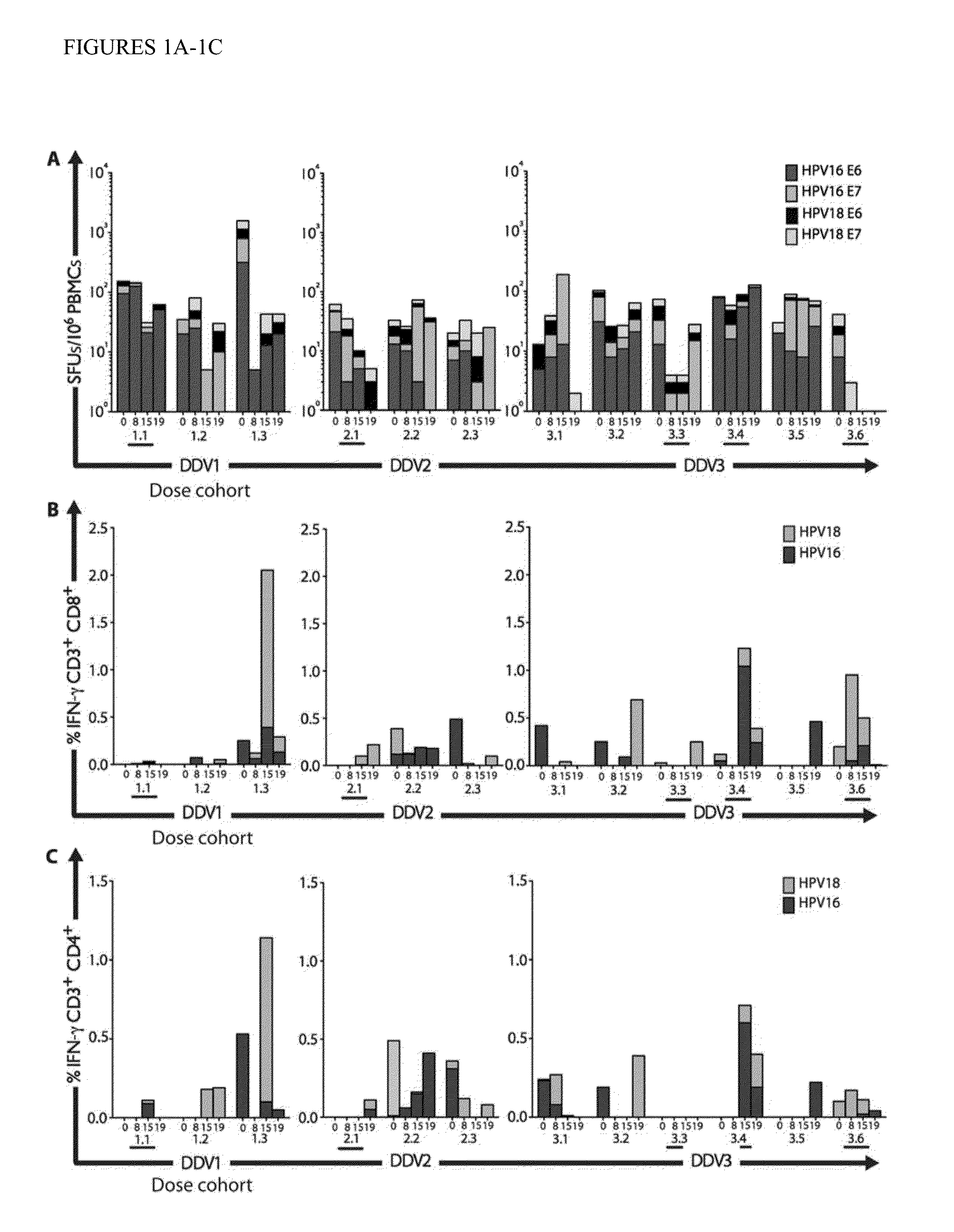
[0101]Therapeutic vaccination elicits HPV-specific cellular immune responses in the blood.

[0102]Standard interferon-γ (IFN-γ) enzyme-linked immunospot (ELISpot) assays (12, 13) were performed to determine the number of antigen-specific IFN-γ-secreting cells in response to stimulation with HPV16 or HPV18 E6 and E7 peptide pools. As shown in FIG. 1, after three vaccinations, one of three subjects who received the low-dose boost vaccination, all three subjects who received the intermediate-dose boost vaccination, and three of six subjects who received the full-dose boost vaccination mounted vaccine-induced HPV16-specific cellular immune responses. Overall, seven subjects (58%) developed cellular immune responses detectable by ELISpot. Peak responses were modest, in the range of 50 to 150 spot-forming units (SFUs) per 106 PBMCs. However, using this assay, increased responses were restricted to HPV16 E7, suggesting that the DNA vaccine, which targeted only the HPV 16 E7 antigen, might in...

example 3

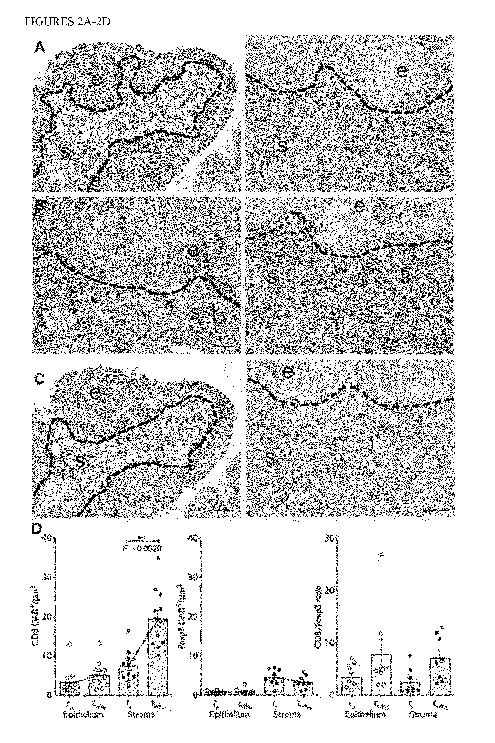
[0104]Tissue CD8+ T cell infiltrates in the target lesion increase after vaccination.

[0105]In unvaccinated persistent CIN2 / 3, we and others have found that tissue T cells are relatively restricted to the stromal compartment immediately subjacent to dysplastic epithelium (11, 14). This pattern of T cell distribution in unvaccinated lesions is consistent with non-antigen-specific recruitment to virally infected mucosa via a chemokine gradient. In contrast, in the post vaccination tissue specimens, the intensity of CD8+ T cell infiltration into lesions increased markedly both in the dysplastic epithelium and in the underlying stromal compartment (P=0.0020) (FIG. 2). These infiltrates were localized in foci of residual dysplasia, not in immediately adjacent normal mucosa. Within-subject increases in tissue CD8+ T cells were significantly greater than the increases we have reported previously in unvaccinated subjects followed over the same time frame (P=0.0300, FIG. 8) (11). These infilt...


PUM

PropertyMeasurementUnit
Timeaaaaaaaaaa
Timeaaaaaaaaaa
Timeaaaaaaaaaa

Abstract

The present invention provides evidence of post-vaccination immunologic changes in target HPV induced cervical intraepithelial neoplasias that suggest the induction of clinically relevant tissue-localized immune responses, despite modest detectable responses in circulating T lymphocytes. The inventive methods allow for identification of immunological responses to HPV immunotherapies, and means for monitoring responses during clinical trials and treatment regimens.

Description

REFERENCE TO RELATED APPLICATIONS[0001]This application claims the benefit of U.S. Provisional Patent Application No. 61 / 919,399, filed on Dec. 20, 2013, which is hereby incorporated by reference for all purposes as if fully set forth herein.STATEMENT OF GOVERNMENTAL INTEREST[0002]This invention was made with U.S. Government support under grant nods. P50 CA098252, 1R21CA123876, R01 CA142691, and 5P30-CA6973 from the National Institutes of Health. The U.S. Government has certain rights in the invention.INCORPORATION-BY-REFERENCE OF MATERIAL SUBMITTED ELECTRONICALLY[0003]The instant application contains a Sequence Listing which has been submitted in ASCII format via EFS-Web and is hereby incorporated by reference in its entirety. Said ASCII copy, created on Dec. 19, 2014, is named P12894-02_ST25.txt and is 3,101 bytes in size.BACKGROUND OF THE INVENTION[0004]On a global scale, human papillomavirus (HPV), most commonly serotype 16 (HPV16), causes about 30% of cancers attributable to in...

Claims


Application Information

Patent Timeline
IPC IPC(8): G01N33/569C12N7/00A61K39/12
CPCG01N33/56972A61K39/12C12N2710/20034A61K2039/585C12N7/00A61K2039/53A61K2039/545A61K2039/55A61K2039/572C12N2710/24143G01N33/57411G01N2333/70517G01N2800/52
Inventor TRIMBLE, CORNELIA
Owner THE JOHN HOPKINS UNIV SCHOOL OF MEDICINE
Why Eureka
Try Eureka