System and method for tracking completeness of co-registered medical image data

a technology of medical image data and completeness, applied in the field of medical imaging, can solve the problems of missing suspicious lesion, difficult to distinguish from other structures or artifacts in the same region, and difficult to find small tumors in the patient's body, and achieve the effect of preventing missing lesions

Active Publication Date: 2015-07-02
METRITRACK
View PDF10 Cites 102 Cited by
  • Summary
  • Abstract
  • Description
  • Claims
  • Application Information

AI Technical Summary

Benefits of technology

[0014]Further, it would be desirable to record inter-image spacing of the still, sequential multiple 2D or 3D images acquired during a particular examination so that the information would be available at a later time for interpretation and also detect, map, and record portions of the region of interest with suboptimal to allow the operator to rescan these regions and, therefore, prevent missing lesions.

Problems solved by technology

However, small tumors are difficult to find in a patient's body and difficult to differentiate from other structures or artifacts in the same region.
Many times a suspicious small finding can coexist in the same region with multiple benign findings (cysts, solid benign nodules, etc.) with similar appearance, which may create confusion during a follow-up examination and may lead to missing the suspicious lesion.
Currently, ultrasound findings are manually labeled by the operator by manually typing or selecting a graphical sign for the current position and orientation of the ultrasound probe and the approximate position of a suspicious lesion in the organ or part of the body, which is time consuming and prone to errors.
Although ultrasound guidance systems and devices do exist, known systems do not offer a practical and accurate solution to mapping targets in 2D or 3D images with real time correction for movement of the patient's body between images and motion of deformable tissue between images.
However, since most ultrasound procedures are manually performed with handheld transducers, the completeness of the scan may be negatively affected by the skill level of the operator or by simple human error.
If the operator fails to maintain the correct transducer speed or orientation during imaging, or if the operator fails to properly follow a given imaging protocol, image data for the complete region of interest may not be acquired.

Method used


Image

  • System and method for tracking completeness of co-registered medical image data
  • System and method for tracking completeness of co-registered medical image data
  • System and method for tracking completeness of co-registered medical image data

Examples


Embodiment Construction

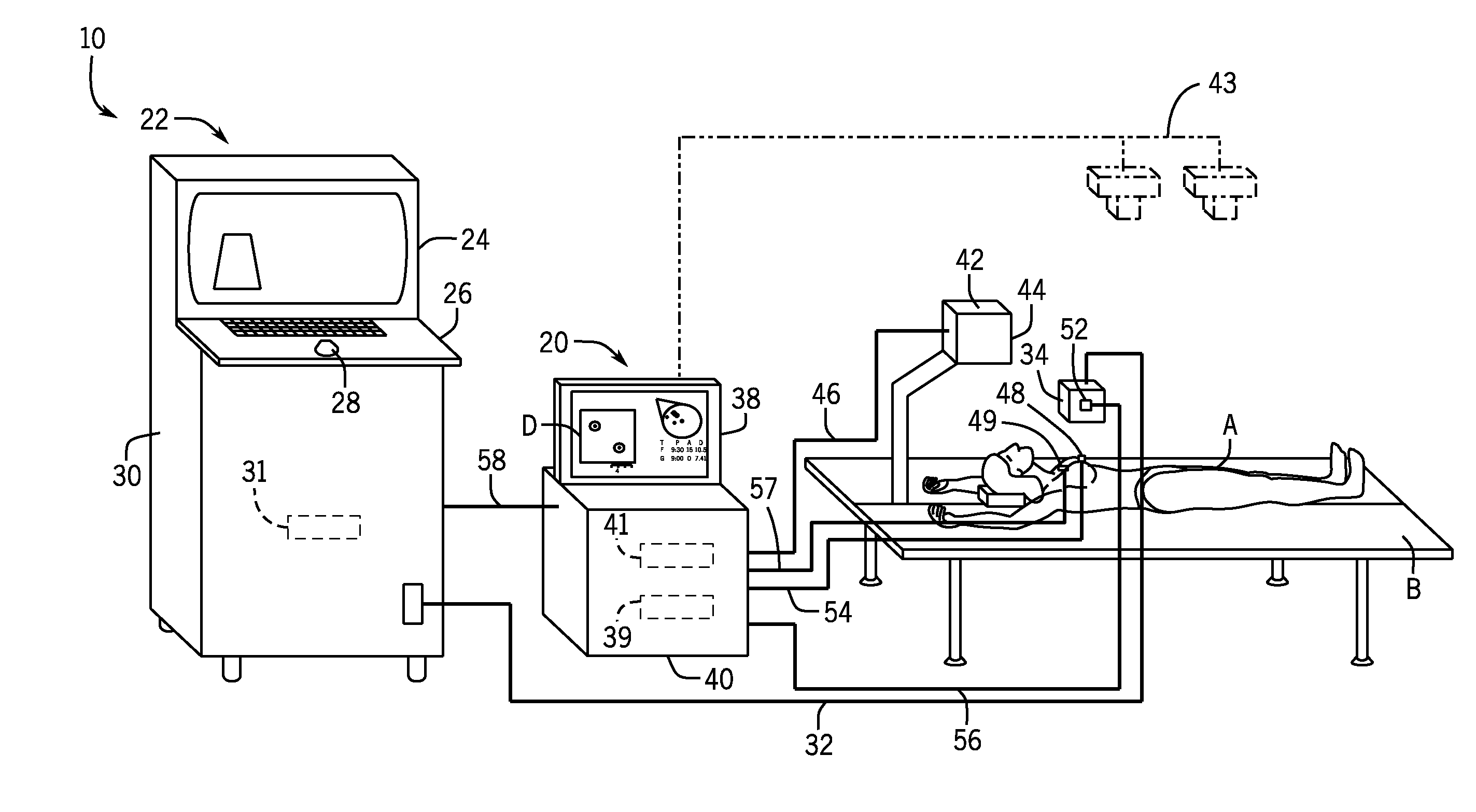
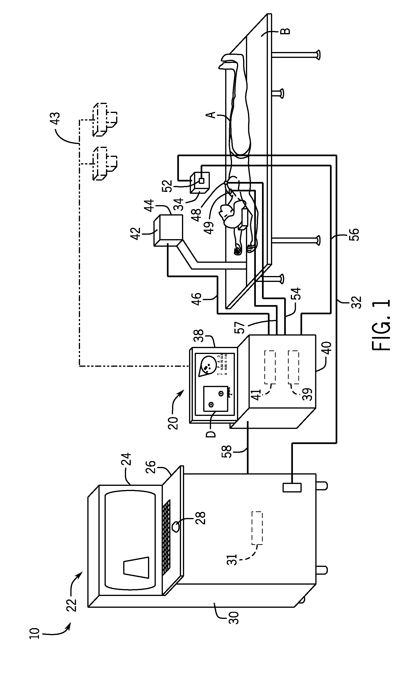
[0046]The operating environment of the various embodiments of the invention are described below with respect to a 2D ultrasound imaging system. However, it will be appreciated by those skilled in the art that the invention the concepts disclosed herein may be extended to 3D ultrasound imaging systems as well as images obtained with a different imaging modality or combination of imaging modalities, such as, for example, x-ray, CT or MRI. Images separately acquired using any of these modalities may be co-registered in space with positional registration to the same anatomical sensor(s) or marker(s) and displayed in a similar manner as described below for ultrasound images. Further, embodiments of the invention may be used for ultrasound breast cancer screening or diagnostic breast ultrasound exams. Additionally, the techniques disclosed herein may be extended to image data acquired from other regions in the body such as, for example, the eye, liver, abdomen, neck, and kidneys.

[0047]Add...


PUM


Abstract

A system and method for tracking completeness of co-registered medical image data is disclosed herein. The system and method tracks the position of an anatomical reference marker positionable on a patient and an ultrasound probe during an imaging session and co-registers medical images based on positional data received from the anatomical reference marker and the ultrasound probe. Using the co-registered image data, the system and method generates a surface contour of a region of interest (ROI) of the patient, such as a breast. The surface contour is defined to represent an interface between a chest wall structure and tissue of the ROI in a plurality of co-registered medical images. A completeness map of the image data within the defined surface contour during the imaging session is generated and overlaid on a graphic representation of the ROI.

Description

CROSS REFERENCE TO RELATED APPLICATION[0001]The present invention is a Non-Provisional of and claims the benefit of U.S. Provisional Patent Application Ser. No. 61 / 964,338, filed Jan. 2, 2014, the disclosure of which is incorporated herein by reference in its entirety.BACKGROUND OF THE INVENTION[0002]Embodiments of the invention relate generally to medical imaging and, more particularly, to a system and method for system for analyzing image data acquired from an imaging modality, generating a surface contour of a region of interest (ROI), and determining the completeness of the acquired image data within the ROI.[0003]Ultrasound imaging systems transmit sound waves of very high frequency (e.g., 1 MHz to 20 MHz) into the patient's body and the echoes scattered from structures in the patient's body are processed to create and display images and information related to these structures. Ultrasound is an important imaging modality for medical diagnostic purposes and as a guidance tool fo...

Claims


Application Information

Patent Timeline
Patent Type & Authority Applications(United States)
IPC IPC(8): A61B8/00A61B19/00A61B6/00A61B5/055A61B8/14A61B8/08
CPCA61B8/4254A61B8/463A61B8/14A61B8/5238A61B8/0825A61B2019/5466A61B5/055A61B19/54A61B2019/5425A61B2019/5454A61B6/502A61B8/469A61B8/483A61B8/5246A61B2017/00694A61B5/061A61B5/1077A61B5/4312A61B6/5247A61B2034/2055A61B2090/3945A61B2090/364A61B2034/2051A61B2090/378A61B90/39A61B2090/3908A61B2090/3925
Inventor CALUSER, CALINANDREI, SILVIU S.
Owner METRITRACK
Why Eureka
Try Eureka