Ultrasound visible catheter

a visible catheter and ultrasonic technology, applied in the field of catheters and catheter placement, can solve the problems of catheter itself not being visualized, catheter placement is incorrect, catheter materials are not acceptable for ultrasound detection, etc., and achieve the effect of easy visualization by ultrasound

Inactive Publication Date: 2015-08-27
GEORGE WASHINGTON UNIVERSITY
View PDF2 Cites 13 Cited by
  • Summary
  • Abstract
  • Description
  • Claims
  • Application Information

AI Technical Summary

Benefits of technology

[0010]Accordingly, it is an object of the invention to provide a catheter that can be visualized by ultrasound. It is a further object of the invention to provide a catheter with an outer texture that reflects ultrasound so that a medical professional can visually see the catheter as it is being placed in the patient to verify proper placement. Another object of the invention is to provide a catheter (including central and peripheral) that is easily visualized by ultrasound, to enable correct placement to be determined and recorded by ultrasound visualization.
[0011]In accordance with these and other objects of the invention, a catheter is provided for use in arterial and / or venous placement, in conjunction with ultrasound positioning. The catheter itself is made of the same materials, and is sized using the standard gauges, as current existing catheters [1]. However, the catheter has a microtexturing on the outer surface of the catheter (added either during fabrication or post-fabrication). This microtexture efficiently and effectively reflects propagated ultrasound waves in situ, to provide a brighter and clearer image of the catheter when being placed in a patient's vessel, with ultrasound imaging equipment.

Problems solved by technology

Properties of current catheter materials are not acceptable for ultrasound detection.
The guide wire and the introducer needle can be visualized under ultrasound, but the catheter itself cannot be visualized.
Because the catheter cannot be visualized, sometimes catheters are placed incorrectly.
Accidental placement of a catheter in an artery instead of a vein can lead to destruction of tissues receiving blood from that artery due to application of medications that damage arteries and are intended only for use in veins.
With the deep and small nature of the vessels used in the ultrasound guided IV catheter technique it can take more time and attention to place these catheters.
Microtextures have been investigated to enhance or inhibit biological interactions of catheters (e.g. antithrombic possessing enhanced drug eluding capabilities [2] and cell ingrowth inhibition [3]), but no one has employed texturing on vascular catheters to make them more reflective / visible to ultrasound.

Method used


Image

  • Ultrasound visible catheter
  • Ultrasound visible catheter
  • Ultrasound visible catheter

Examples


Embodiment Construction

[0029]In describing a preferred embodiment of the invention illustrated in the drawings, specific terminology will be resorted to for the sake of clarity. However, the invention is not intended to be limited to the specific terms so selected, and it is to be understood that each specific term includes all technical equivalents that operate in similar manner to accomplish a similar purpose. Several preferred embodiments of the invention are described for illustrative purposes, it being understood that the invention may be embodied in other forms not specifically shown in the drawings.

[0030]Reflective Patterns

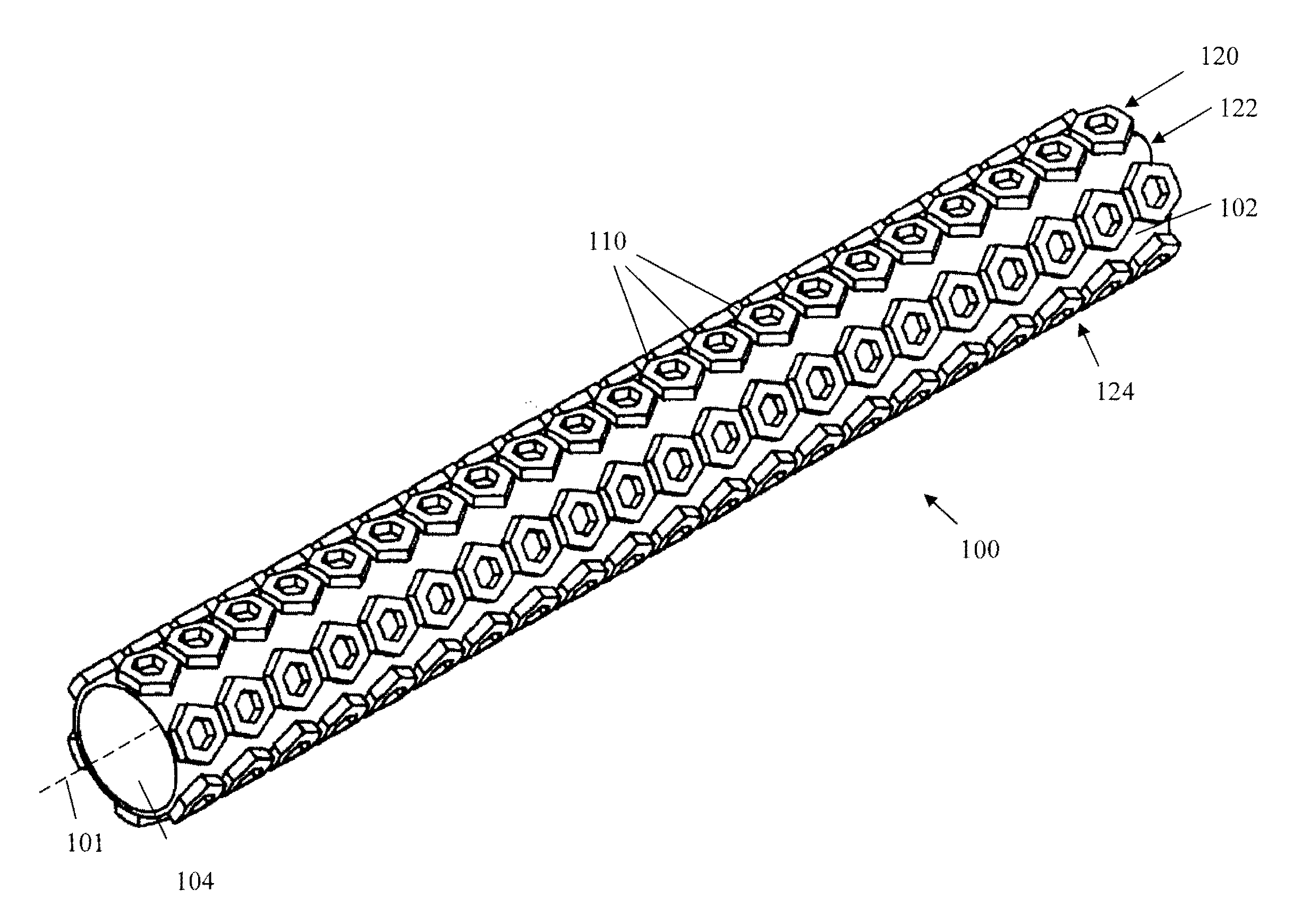
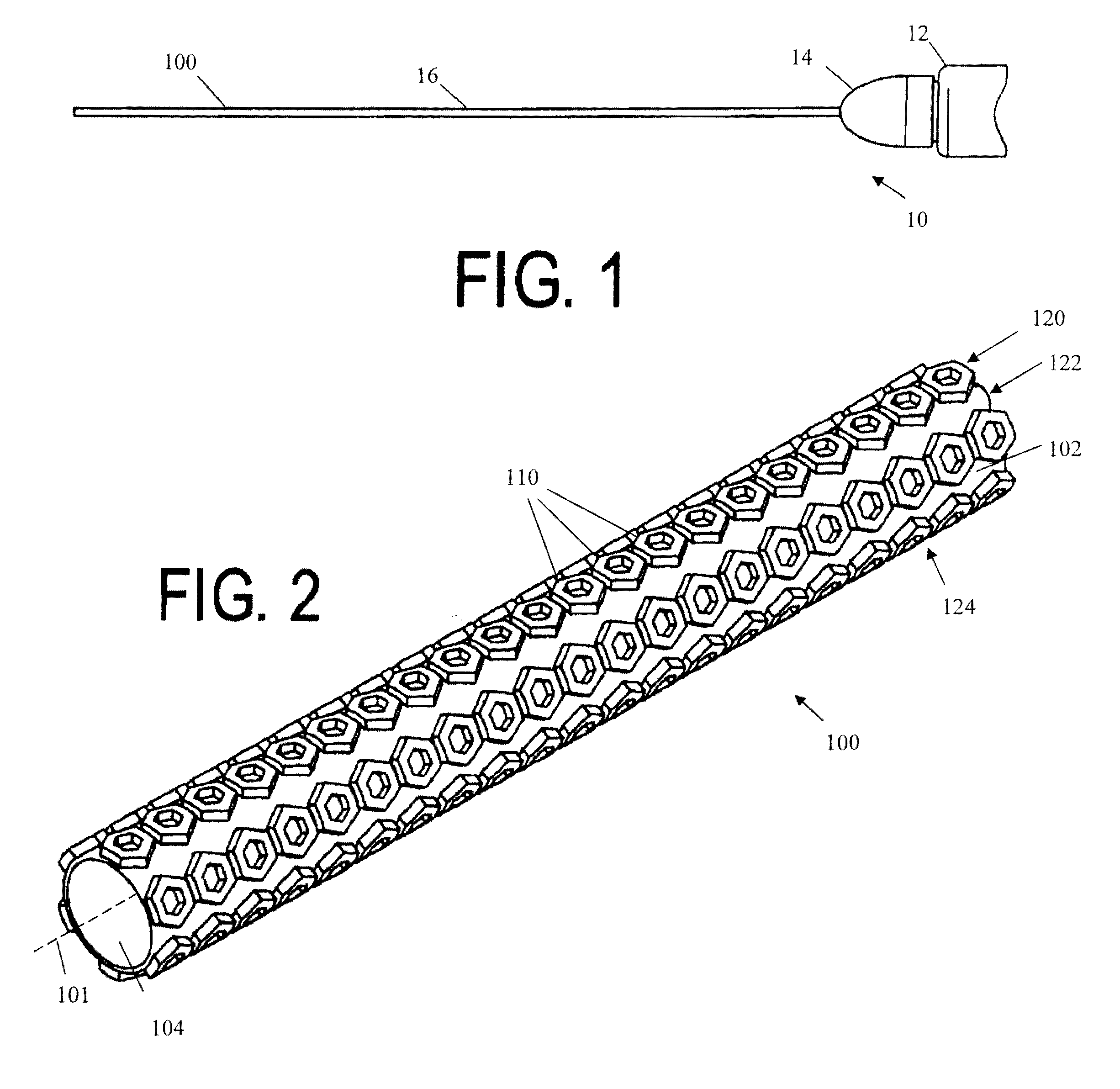
[0031]Referring to the drawings, FIG. 1 shows the catheter / needle assembly 10 of the present invention. The assembly 10 includes a round tubular housing 12, hub 14, needle 16 and catheter 100. The housing 12 has a first end that is open and can mate with a medication container or drain. The needle 16 can extend through openings in the hub 14 and housing 12. The catheter 100 exten...


PUM


Abstract

A catheter can be used in arterial and / or venous placement, in conjunction with ultrasound positioning. The catheter itself is made of the same materials, and is sized using the standard gauges, as current existing catheters. However, the catheter has a microtexture on the outer surface of the catheter. This microtexture efficiently and effectively reflects propagated ultrasound waves in situ, to provide a brighter and clearer image of the catheter when being placed in a patient's vessel, with ultrasound imaging equipment.

Description

RELATED APPLICATIONS[0001]This application is a continuation-in-part of PCT / US2014 / 018931, filed Feb. 27, 2014, which claims benefit of U.S. Provisional Application No. 61 / 770,052, filed Feb. 27, 2013. This application also claims the benefit of U.S. Provisional Application No. 61 / 945,611, filed Feb. 27, 2014. The entire contents of each of those applications are incorporated herein by reference.BACKGROUND OF THE INVENTION[0002]1. Field of the Invention[0003]The present invention relates to catheters and the placement of catheters in a patient. More particularly, the present invention relates to a catheter that is visible to ultrasound to assist placement of the catheter in a patient.[0004]2. Background of the Related Art[0005]Catheters are small tubes that can be inserted into the body for medical purposes, such as to introduce or remove a gas or fluid for the treatment of an illness or disease or to perform a surgical procedure. The process of inserting a catheter into a body cavi...

Claims


Application Information

Patent Timeline
Patent Type & Authority Applications(United States)
IPC IPC(8): A61M25/01A61B8/08
CPCA61B8/0841A61M25/0108A61M2025/006A61B5/061A61B90/39
Inventor CORMAN, ADAMSIKKA, NEAL K.CASTRO, NATHAN J.HOLMES, BENJAMINALMANSOURI, HAWAA
Owner GEORGE WASHINGTON UNIVERSITY
Why Eureka
Try Eureka