Non-Invasive Imager for Medical Applications

Active Publication Date: 2015-09-10
THOMAS MAMMEN +1
View PDF4 Cites 18 Cited by
  • Summary
  • Abstract
  • Description
  • Claims
  • Application Information

AI Technical Summary

Benefits of technology

[0005]The present invention is aimed at providing the medical and veterinary community tools to conduct invasive procedures with visibility and control using spatially aligned holographic imaging in real-time for guidance of tools and placement of medicines while enabling real-time viewing and follow-up by the surgical team and experts during the procedure itself. The invention is also an enabler using the 3-D image for improving the diagnosis of proble

Problems solved by technology

Computed Tomography (CT) scan that use either X-rays or Gamma rays, and standard X-ray imaging, though much have improved over the past years to provide good images using lower radiation levels, still damage the tissues due to accumulated radiation dosage.
These high resolution scans require mathematical computations that take time and hence cannot be don

Method used


Image

  • Non-Invasive Imager for Medical Applications
  • Non-Invasive Imager for Medical Applications
  • Non-Invasive Imager for Medical Applications

Examples


Embodiment Construction

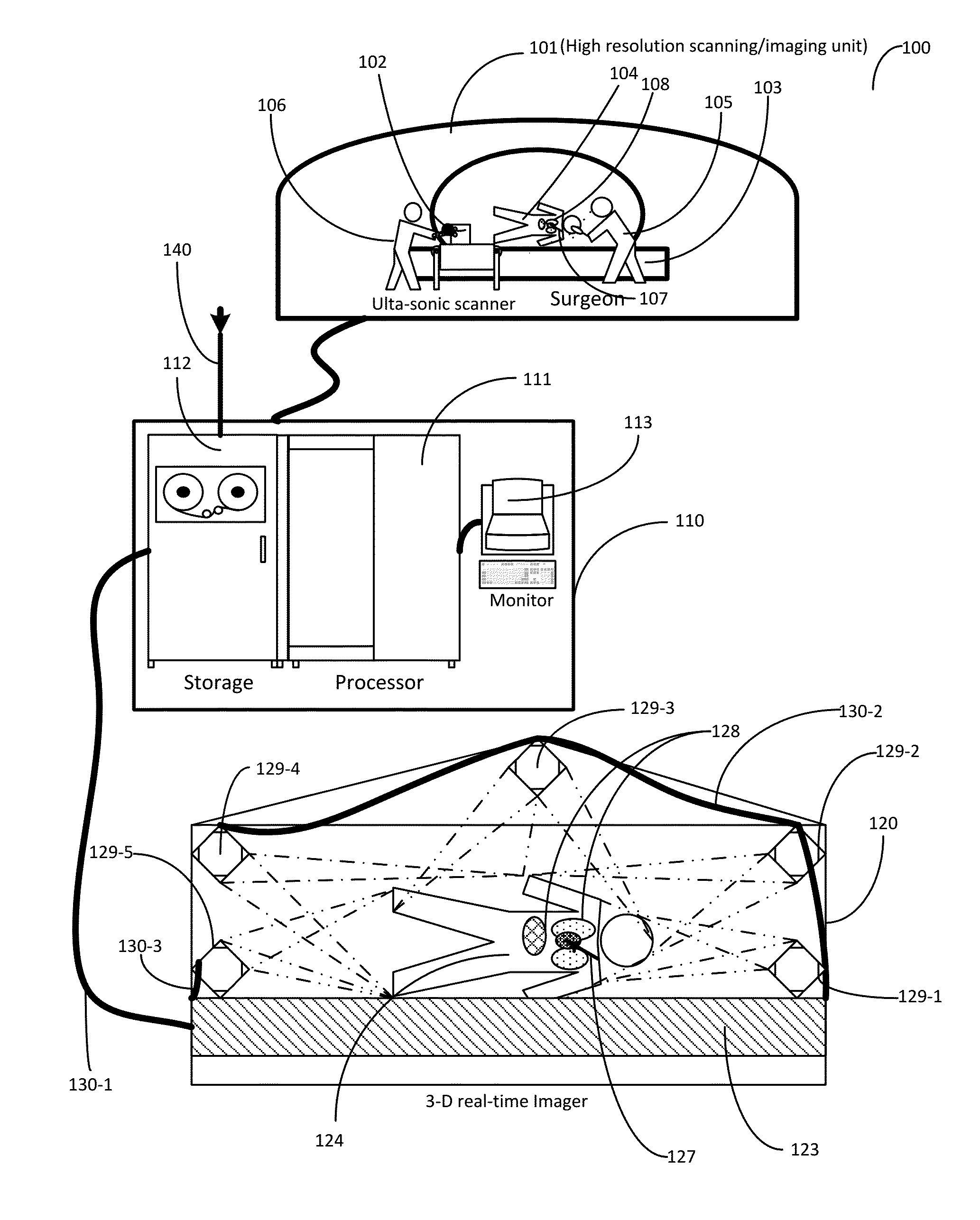
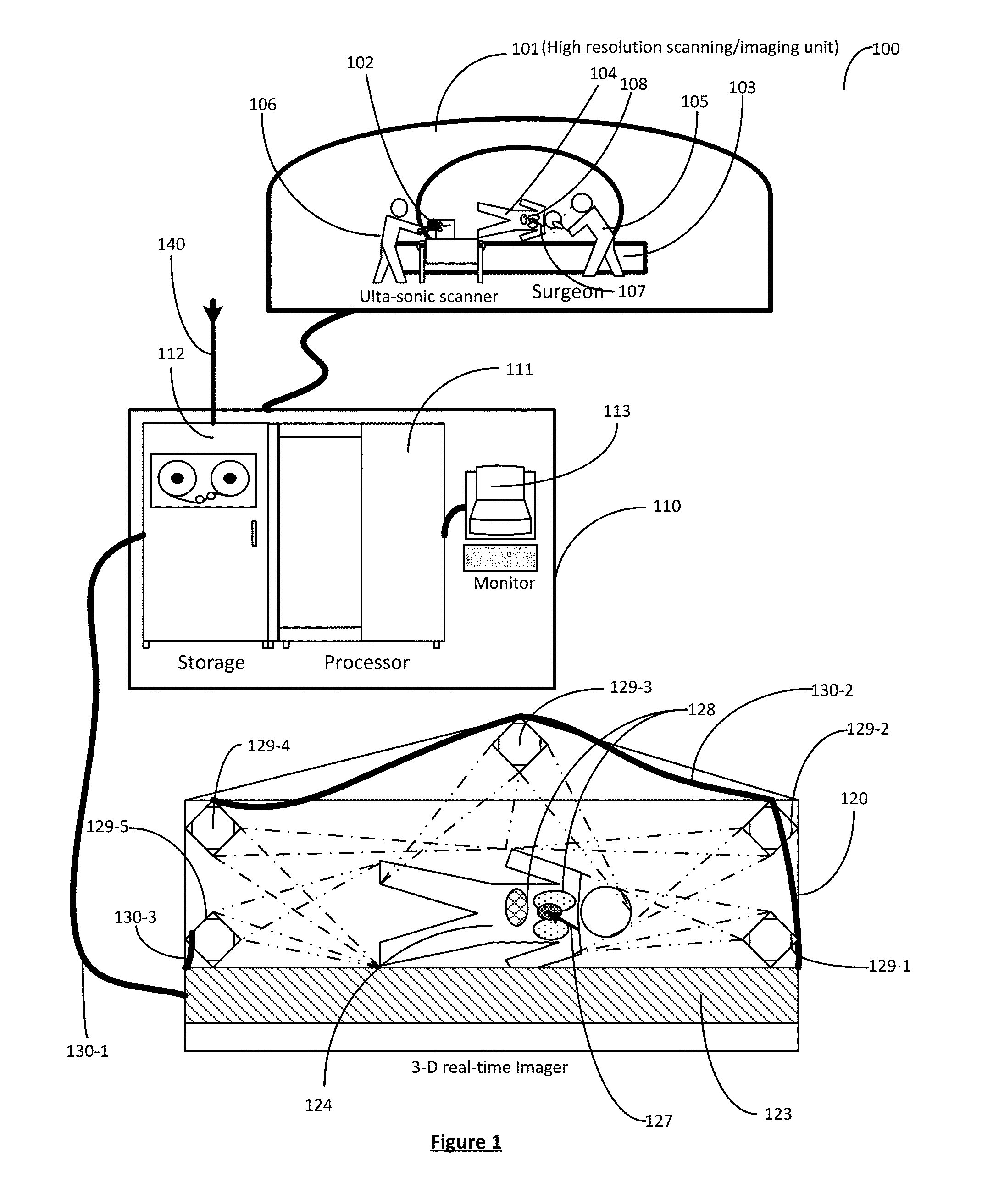
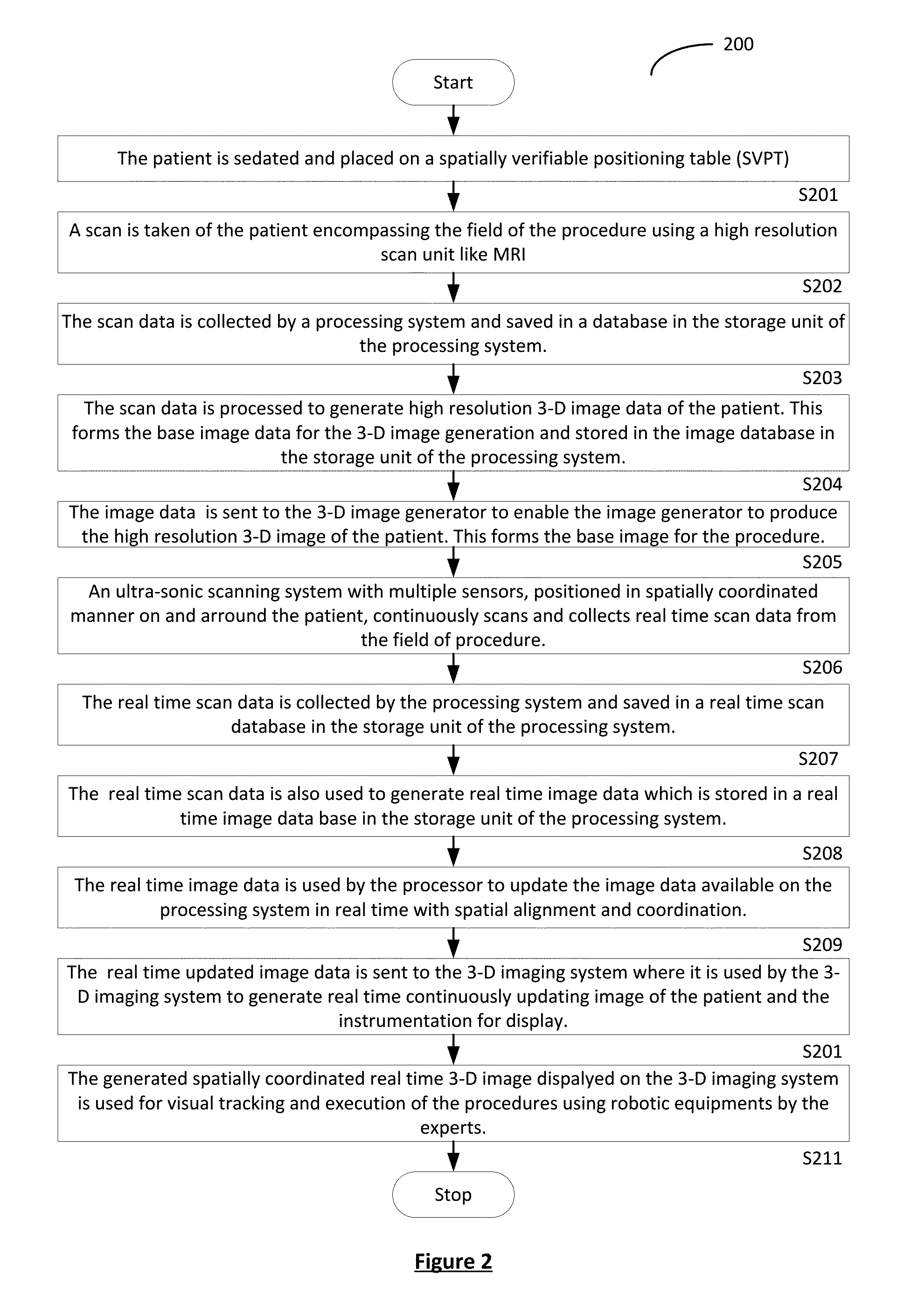
[0025]A method and process is described for providing Non-Invasive3-D image (e.g. a three dimensional holographic image) of the patient in a spatially coordinated and updatable manner, such that during surgical or other procedures the person performing the procedure can visually identify the organs and the location of the instruments in real time inside the body. Hence a spatially aligned non-invasive imaging and reconstruction using any available 3-D image generator, such as a 3-D projector, generating a 3-D image of the patient, will be very valuable tool to the surgical community. The high powered computing capabilities, advances in the imaging techniques, individually or in combination, when combined with noise filtering and error correction capabilities, have made accurate 3-D imaging such as 3-D holograms from scans a reality. These 3-D images are usable as a diagnostic tool and implementation tool by the medical community. It can also be a valuable teaching tool. There may be...


PUM


Abstract

A method and process is described for providing Non-Invasive three dimensional (3-D) image of a patient such that during surgical or other procedures, the person performing the procedure can visually identify the organs and the location of the instruments in real time inside the body. Such a non-invasive imaging and reconstruction using spatially coordinated imaging in three dimensions is a very valuable tool especially to the surgical community. The high powered computing capabilities, advances in the imaging techniques, individually or in combination when combined with noise filtering and error correction capabilities, have made accurate 3-D imaging in real time from scans a reality. These 3-D images are also used as a diagnostic tool, a practice tool and a teaching tool by the medical community. There may be other applications in these and related areas which may emerge as technology develops and they become apparent to individuals practicing the art.

Description

FIELD OF INVENTION[0001]This invention addresses the need of the medical and veterinary community to visualize in real time and in three dimensions, the field of operation and the location of instruments during procedures and also accurate placement of medicines.DESCRIPTION OF BACKGROUND OF THE INVENTION[0002]Today most of the robot assisted procedures are conducted by the surgeon operating with a limited vision of the operating field, through microscopes and manipulating the instruments by what is visible in the narrow field of view through the optical cable inserted into the body with the surgical instrumentation. The surgeon has no real wide view of the operating area. There has been scanning and imaging technology such as magnetic resonance imaging (MRI), computed tomography (CT), X-ray, ultra-sound (ultrasonic imaging) etc. which are in wide spread use in diagnostic fields. 3-D imaging technology is also available today, to a limited extend using the images generated which are ...

Claims


Application Information

Patent Timeline
IPC IPC(8): A61B8/00A61B5/055A61B5/00A61B8/08
CPCA61B8/4416A61B5/72A61B5/055A61B8/483A61B5/062A61B5/7425A61B8/0841A61B8/085A61B2090/364A61B2090/367
Inventor THOMAS, MAMMENTHOMAS, ARUN MAMMEN
Owner THOMAS MAMMEN
Why Eureka
Try Eureka