Human endocrine progenitors from adult pancreatic tissue

Inactive Publication Date: 2015-09-24
THE JOHN HOPKINS UNIV SCHOOL OF MEDICINE
View PDF0 Cites 4 Cited by
  • Summary
  • Abstract
  • Description
  • Claims
  • Application Information

AI Technical Summary

Benefits of technology

[0006]As described herein, the present inventors demonstrate the presence of NGN3 protein-expressing cells in histologically normal duct and acinar cells in the adult human pancreas. NGN3+ cells accumulate following short-term culture of pancreatic tissue. The NGN3+ cell population is negatively regulated by Notch signaling and proteasome degradation and positively regulated by EGF receptor and STAT3 signaling. NGN3+ cells have high aldehyde dehydrogenase (ALDH) activity and coexpress the cell surface g

Problems solved by technology

Even the most diligent patients suf

Method used


Image

  • Human endocrine progenitors from adult pancreatic tissue
  • Human endocrine progenitors from adult pancreatic tissue
  • Human endocrine progenitors from adult pancreatic tissue

Examples


Example

Example 1

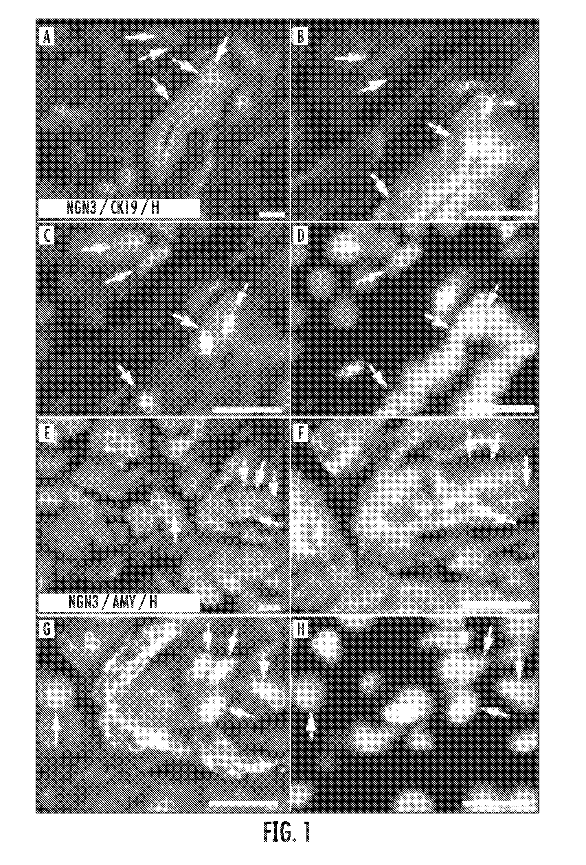
NGN3 is Expressed by Acinar and Duct Cells in the Adult Human Pancreas

[0091]NGN3 expression was detected in grossly and histologically normal tissues from five surgically resected pancreata. In all five pancreata, NGN3 was localized in the nucleus of amylase (AMY)+ acinar cells and cytokeratin 19 (CK19)+ duct cells (FIG. 1A-H). The level of NGN3 expression varied between patients and was both heterogeneous and spatially restricted within individual tissue sections. In randomly selected fields of >100 nuclei, the mean±s.e.m % NGN3+ cells was 2.4±1.1%. Exocrine NGN3 expression was identified in an additional 15 patient biopsies.

Example

Example 2

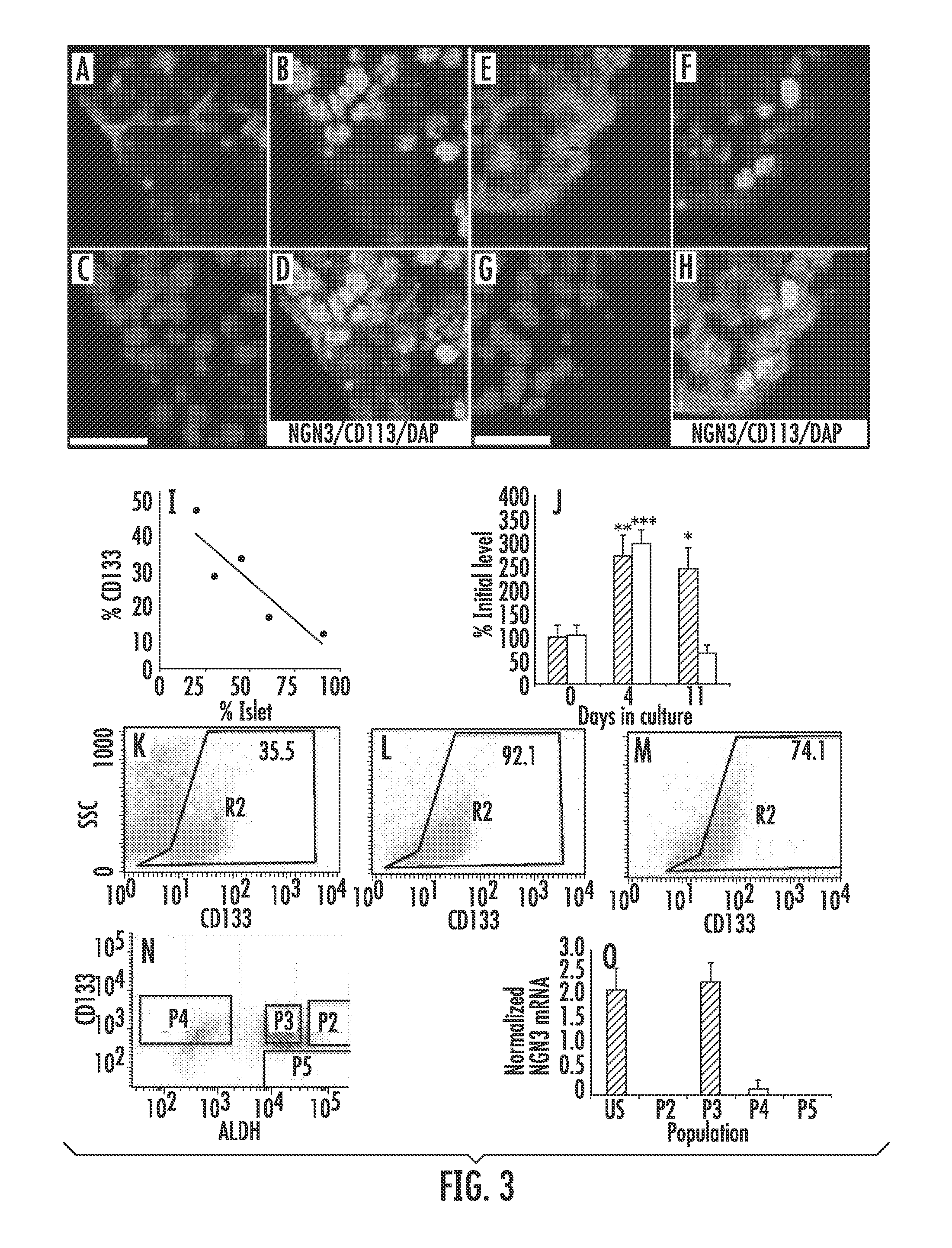
NGN3 Protein and Neurogenic Differentiation 1 (NEUROD1) mRNA Expression Increases During Culture of Pancreatic Tissue

[0092]Enzymatically-digested adult human cadaveric pancreas preparations containing a mixture of exocrine tissue and islets (pancreatic tissue) were received two to three days post mortem (D2-D3) and cultured in defined media for 4 days. This culture system was used to investigate changes in the expression of NGN3 in tissue undergoing ADM. In three independent (different organ donors) pancreatic tissue preparations, the mean±s.e.m percentage of cells expressing AMY on D7 decreased significantly to 7.3%±3.4 relative to D3 (P<0.001, n=3). The mean±s.e.m percentage of cells expressing CK19 relative to D3 increased significantly to 180.7%±19.6 (P<0.01, n=3).

[0093]The percentage of cells expressing nuclear NGN3 protein (representative images of NGN3 expression in cultured tissue in FIG. 3B, F) increased significantly after 4 days in culture compared to initial lev...

Example

Example 3

NGN3 is Negatively Regulated by Notch Signaling

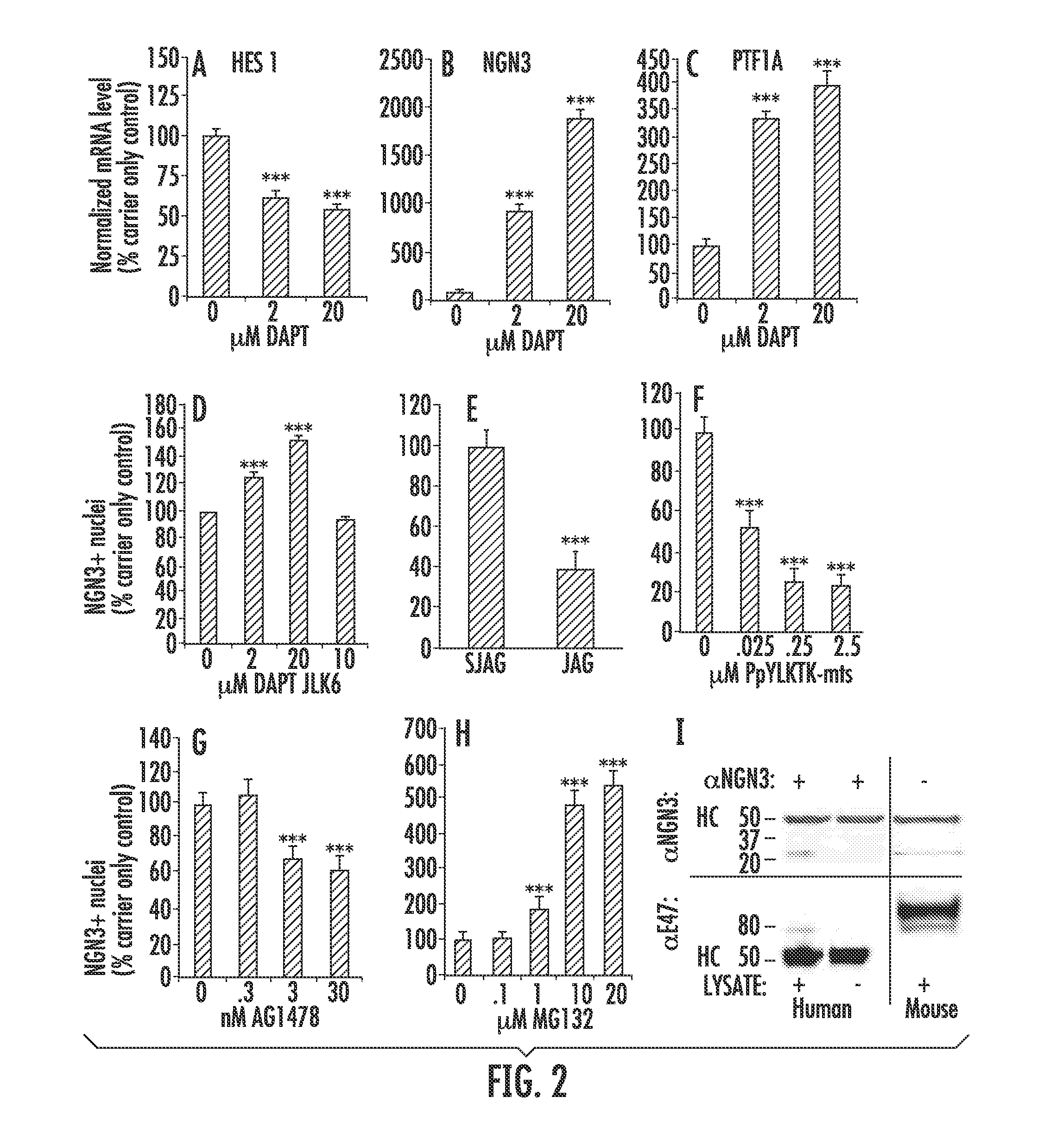
[0094]In order to investigate the involvement of Notch signaling in cultured pancreatic tissue, Notch was inhibited with the γ-secretase inhibitor DAPT (25) and the mRNA levels of the Notch downstream negative regulator of NGN3, Hairy and Enhancer of Split 1 (HES1) (1, 26, 27) were determined as was the mRNA and protein levels of NGN3. The level of HES1 mRNA decreased significantly after 4 days of culture in the presence of 2 μM and 20 μM DAPT to 63.0%±3.3 (P<0.001, n=6 readings) and 55.4%±1.5 (P<0.001, n=6 readings) of carrier control, respectively (FIG. 2A). The level of NGN3 mRNA increased significantly in the presence of 2 μM and 20 μM DAPT to 927.4%±52.6 (P<0.001, n=6) and 1886.3%±102.4 (P<0.001, n=6) of carrier control, respectively (FIG. 2B).

[0095]PTF1A plays a critical role in the formation and spatial organization of the murine exocrine and endocrine pancreas (28) and marks precursor cells that give rise to all exocrin...


PUM


Abstract

The present invention relates to the field of progenitor cells. More specifically, the present invention provides compositions and methods for isolating endocrine progenitor cells from pancreatic tissue. In certain embodiments, the method comprises the steps of (a) providing a pancreatic tissue sample; (b) isolating cells positive for CD133+; and (c) culturing the isolated cells in defined media for at least about 4 days.

Description

CROSS-REFERENCE TO RELATED APPLICATIONS[0001]This application claims the benefit of U.S. Provisional Application No. 61 / 712,991, filed Oct. 12, 2012; which is incorporated herein by reference in its entirety.FIELD OF THE INVENTION[0002]The present invention relates to the field of progenitor cells. More specifically, the present invention relates to the isolation of human endocrine progenitor cells.BACKGROUND OF THE INVENTION[0003]Patients with type I (juvenile) diabetes lack pancreatic beta cells. Beta cells produce insulin, the hormone responsible for glucose homeostasis. In the absence of beta cells, type I diabetics must mechanically test and administer insulin to stay alive. Even the most diligent patients suffer significant lifetime morbidity. A great need exists for alternative methods and compositions for treating type I diabetics.SUMMARY OF THE INVENTION[0004]The present invention is based, at least in part, on the discovery that human endocrine progenitor cells can be isol...

Claims


Application Information

Patent Timeline
IPC IPC(8): A61K35/39C12N5/071A61K35/55G01N33/569
CPCA61K35/39G01N33/56966C12N5/0613A61K35/55C12N5/0676C12N2506/07G01N2333/4703G01N2405/10C12N2533/90C12N2533/40G01N2333/70596C12N5/0678G01N33/507G01N33/5073G01N2800/042
Inventor SHAMBLOTT, MICHAEL J.
Owner THE JOHN HOPKINS UNIV SCHOOL OF MEDICINE
Why Eureka
Try Eureka