System and method for diagnosis of focal cortical dysplasia

a cortical dysplasia and automatic detection technology, applied in the field of medical imaging, can solve the problems of affecting the quality of life of patients, and causing a large amount of data, so as to achieve accurate registrization of the cerebral hemisphere, automatic detection and localization of focal cortical dysplasia, and the effect of limiting the amount of data

Inactive Publication Date: 2015-10-15
THE GENERAL HOSPITAL CORP
View PDF2 Cites 15 Cited by
  • Summary
  • Abstract
  • Description
  • Claims
  • Application Information

AI Technical Summary

Benefits of technology

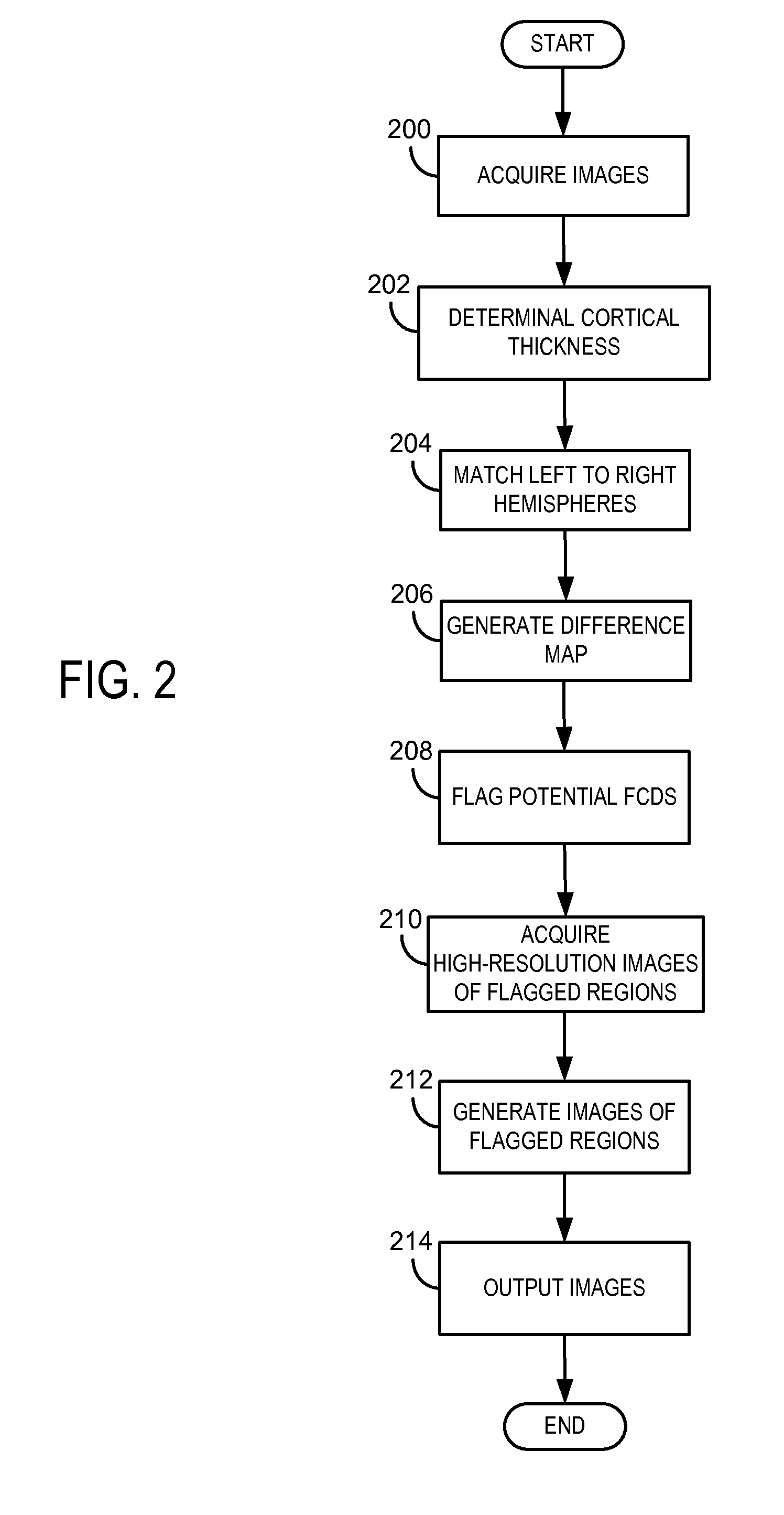
[0012]The present invention overcomes the aforementioned drawbacks by providing a system and method for automatically detecting and localizing focal cortical dysplasias. The invention can be used to accurately register the cerebral hemisphere on one side of the brain to the hemisphere on the other side. The present invention recognizes that corresponding locations have approximately the same thickness, except for regions that have dysplasias. Following identification of these regions, high spatial resolution data is acquired only of these regions so that high resolution images of the regions can be displayed for manual examination. As the output images only include regions of potential dysplasias rather than the whole brain, this invention dramatically limits the amount of data that a neuroradiologist must view in order to make a diagnosis.

Problems solved by technology

Epilepsy, a common neurological disorder characterized by recurrent unprovoked seizures, exacts a large toll upon society in terms of both quality of life and health care costs.
Furthermore, the morbidity of epilepsy is great, in part because epilepsy, unlike many other neurologic disorders, affects patients of all ages and can significantly impair a patient's quality of life for many years.
Indeed, the incidence of new cases of epilepsy is seen in the first year of life, thus accounting for the cost-intensive nature of the disorder.
These are subtle variations in the thickness and signal characteristics in the brain's cerebral cortex, a structure that is so highly convoluted and anatomically irregular that it is difficult for the human eye to detect small abnormalities, thus making FCD often very difficult to detect by even the most experienced subspecialist neuroradiologists.
For example, during visual analysis of MRI images, the foldings of the cortex make diagnosis exceedingly difficult as a visual estimation of the thickness (defined as the distance between the gray / white boundary and the pial surface) will invariably be inaccurate in regions where the surfaces are not parallel to either each other or one of the cardinal imaging planes.
Unfortunately, images acquired at this resolution across the entire brain represent an enormous amount of data for a radiologist to examine in order to detect a subtle abnormality.
Furthermore, merely screening for the general location of an abnormality is insufficient.
As discussed above, high-resolution MRI places a great burden on neuroradiologists as they must scan through hundreds or thousands of slices in order to detect the subtle FLAIR brightening (the hallmark of FTDs) on only a few images.
While these approaches can exhibit good sensitivity in homogeneous, controlled research studies, they are likely to fail to detect FTDs in practice.
More specifically, since small FTDs that are difficult to diagnose frequently present without detectable focal cortical thickening, typical “thickness-detection” approaches for diagnosing cannot be used.
Furthermore, analyzing FLAIR images for small changes in signal intensities is a very time-consuming, and thus impractical, approach.

Method used


Image

  • System and method for diagnosis of focal cortical dysplasia
  • System and method for diagnosis of focal cortical dysplasia
  • System and method for diagnosis of focal cortical dysplasia

Examples


example

[0065]The above processing methods and techniques were applied in a study examining the feasibility of aspects of the present invention. The study included six patients with post-operative diagnosis of FCDs, but a “negative” diagnosis from conventional MRI procedures and examination. In the study, MRI images were analyzed in accordance with methods of the present invention described above, and all six FTDs previously missed on clinical reads were detected, with an average of more than 15 years of potentially treatable seizures. The results of the study therefore indicated that the methods of the present invention can help identify possible FTDs in cases in which the dysplasias would otherwise have gone undetected, resulting in decades of potentially treatable seizures. The following paragraphs further describe materials, methods, and results of the study.

[0066]The six subjects used in the study were identified as having surgery for FCDs that carried a post-operative diagnosis of FTD...


PUM


Abstract

A system and method for automatic detection of potential focal cortical dysplasias through magnetic resonance imaging. The method includes acquiring image data of a subject brain at a first resolution, analyzing the acquired image data to determine a thickness of cerebral gray matter, and matching the left cerebral hemisphere to the right cerebral hemisphere based on corresponding geometric features of the hemispheres. The method also includes generating a difference map comparing corresponding thicknesses of the hemispheres, identifying regions of abnormal differences in thickness as potential regions containing focal cortical dysplasias, and acquiring image data of the regions of abnormal differences in thickness at a second resolution. The method further includes generating images of the regions of abnormal differences in thickness from the acquired image data and displaying the images.

Description

CROSS-REFERENCE TO RELATED APPLICATIONS[0001]This application is based on, claims priority to, and incorporates herein by reference in its entirety U.S. Provisional Application Ser. No. 61 / 715,779, filed Oct. 18, 2013, and entitled “SYSTEM AND METHOD FOR DIAGNOSIS OF FOCAL CORTICAL DYSPLASIA USING MAGNETIC RESONANCE IMAGING.”STATEMENT REGARDING FEDERALLY SPONSORED RESEARCH[0002]This invention was made with government support under NS052585 and P41-RR14075 awarded by the National Institutes of Health. The government has certain rights in the invention.BACKGROUND OF THE INVENTION[0003]The present invention relates generally to systems and methods for medical imaging and, more particularly, the invention relates to systems and methods for automated detection of focal cortical dysplasias in medical images.[0004]Epilepsy, a common neurological disorder characterized by recurrent unprovoked seizures, exacts a large toll upon society in terms of both quality of life and health care costs. ...

Claims


Application Information

Patent Timeline
Patent Type & Authority Applications(United States)
IPC IPC(8): A61B5/055G01R33/385G06K9/46A61B5/107A61B5/00G06T7/00G01R33/54G01R33/34
CPCA61B5/055G06T2207/30016G01R33/385G01R33/34A61B5/1072A61B5/4064A61B5/0046A61B5/0042A61B5/0037G06T7/0012G06T7/0081G06K9/4609A61B2576/026G06K2209/051G06T2207/10088G01R33/543A61B5/1075G01R33/5608G16H30/40G06V2201/031
Inventor FISCHL, BRUCECOPEN, WILLIAM A.
Owner THE GENERAL HOSPITAL CORP
Why Eureka
Try Eureka