Automatic identification of a potential pleural effusion

a technology of pleural effusion and automatic identification, which is applied in the field of automatic identification of potential pleural effusions, can solve the problems of suffocation of patients, inaccurate pleural effusion identification data, and time-consuming determination, just like manual segmentation,

Active Publication Date: 2015-10-22
SIEMENS HEALTHCARE GMBH
View PDF4 Cites 14 Cited by
  • Summary
  • Abstract
  • Description
  • Claims
  • Application Information

AI Technical Summary

Benefits of technology

[0051]Moreover, it is possible to make statements in relation to the location of a potential pleural effusion, namely in particular as to whether a potential pleural effusion is situated exclusively in the right-hand or left-hand thoracic cavity half, or whether it is situated in both halves. This information may be particularly valuable to a treating practitioner because it may allow conclusions to be drawn about the pathological cause of the potential pleural effusion.

Problems solved by technology

Suffocation of the patient may be the result in extreme cases.
However, none of these constants were found to be sufficiently correct, and so this determination, just like the manual segmentation, is not only time-consuming but also, moreover, inaccurate.
If the HU-value in the region of a potential pleural effusion identified by the pleural effusion identification data deviates significantly, the assumption can be made that the pleural effusion identification data are faulty.

Method used


Image

  • Automatic identification of a potential pleural effusion
  • Automatic identification of a potential pleural effusion
  • Automatic identification of a potential pleural effusion

Examples


Embodiment Construction

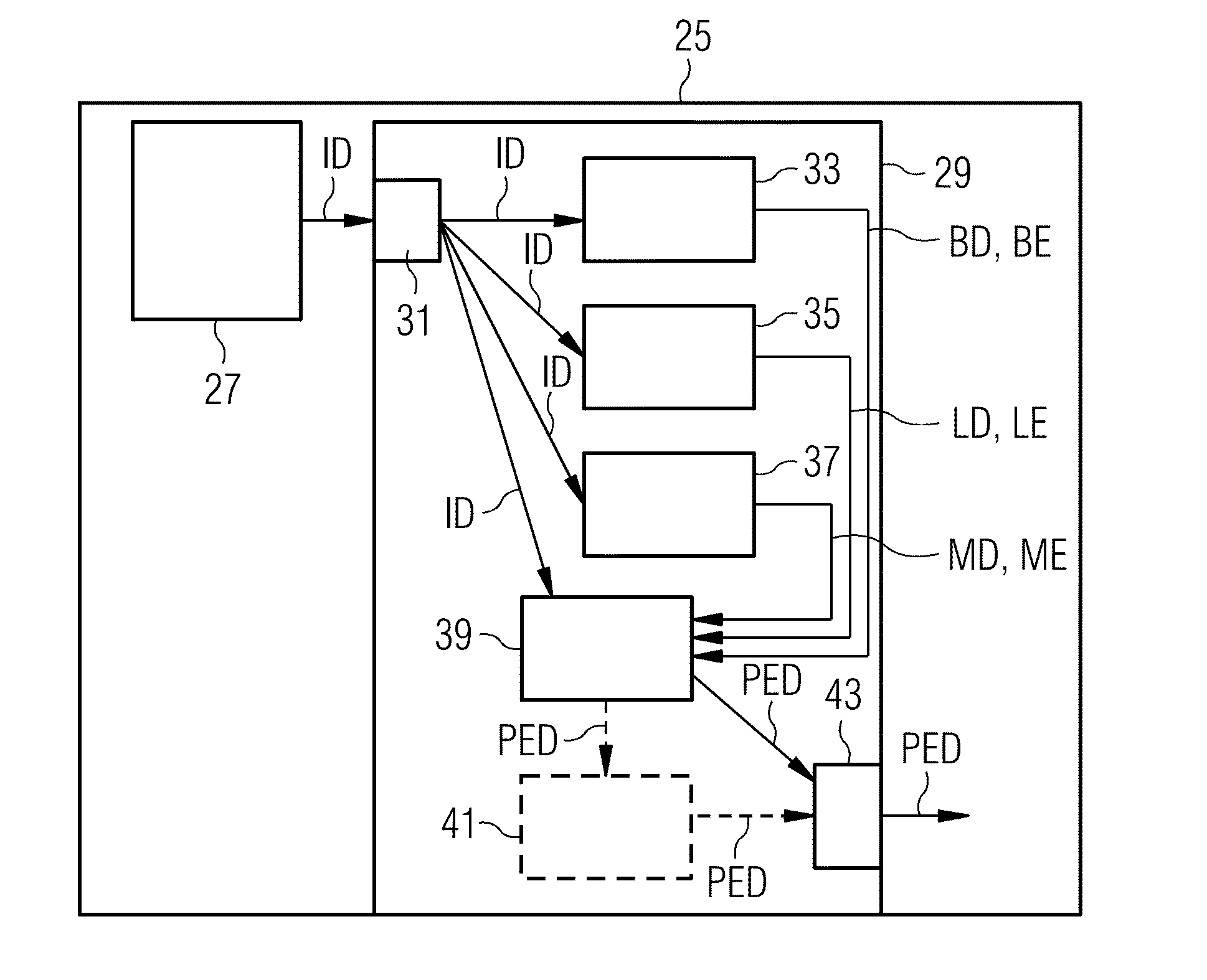
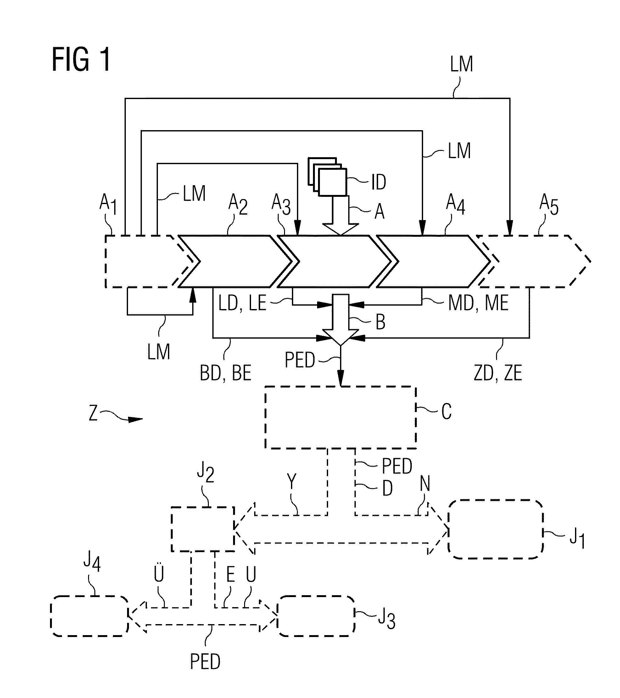
[0063]FIG. 1 shows a schematic flowchart of an exemplary embodiment of the method Z according to embodiments of the invention for identifying a potential pleural effusion. Here, image data ID of a patient are subjected to an automatic identification of a potential pleural effusion in a step A. This step A comprises a plurality of sub-steps A1, A2, A3, A4, A5 (some of which are mandatory, some of which are optional). In a first, optional sub-step A1, there is a landmark detection A1 in the image data ID, with the aid of which landmarks LM are extracted, which landmarks may serve for orientation in the image data ID and, in particular, for segmenting individual organs or structures. A second sub-step A2 is an acceptance A2 of rib cage detection data BD of the rib cage of the patient from the image data. These rib cage detection data BD comprise rib cage extent data BE, i.e. data which represent the rib cage extent of the interior of the rib cage of the patient.

[0064]This acceptance ma...


PUM


Abstract

A method for automatically identifying a potential pleural effusion in medical image data of a thorax of a patient from a scan by means of a medical scanner is provided. It includes at least the steps of accepting rib cage detection data of the rib cage of the patient from the image data, which rib cage detection data include rib cage extent data of a rib cage extent of the interior of the rib cage, accepting lung detection data of the lung of the patient from the image data, which lung detection data comprise lung extent data of a lung extent of the external boundary of the lung, accepting mediastinum detection data of all organs of the mediastinum in the thorax (Th) of the patient from the image data, which mediastinum detection data comprise mediastinum extent data of a mediastinum extent of the external boundary of the mediastinum, and subtracting the lung extent and the mediastinum extent from the rib cage extent while forming pleural effusion identification data.

Description

CROSS REFERENCE TO RELATED APPLICATIONS[0001]This application claims priority to DE 102014207449.5 having a filing date of Apr. 17, 2014, the entire contents of which are hereby incorporated by reference.FIELD OF TECHNOLOGY[0002]The following relates to a method for automatically identifying a potential pleural effusion in medical image data of a thorax of a patient from a scan by means of a medical scanner. It moreover relates to an identification system for such an identification.BACKGROUND[0003]In medical terms, a pleural effusion is an abnormal collection of liquid in the pleural cavity, which is a narrow gap between the pleurae. The liquid therefore is situated in the rib cage between the lung and the ribs.[0004]In healthy humans, the pleural cavities are filled with a small amount—approximately 5 ml—a high-protein liquid, which, as a type of “lubrication layer”, provides freedom of movement to the lungs during inhalation and exhalation.[0005]Smaller pleural effusions (up to a ...

Claims


Application Information

Patent Timeline
Patent Type & Authority Applications(United States)
IPC IPC(8): A61B6/00A61B6/03
CPCA61B6/032A61B6/5217A61B8/5223G06T7/0014G06T2207/10081G06T2207/10132G06T2207/20224G06T2207/30061G16H50/30
Inventor DANKERL, PETERHAMMON, MATTHIASKRAMER, MARTINSOZA, GRZEGORZ
Owner SIEMENS HEALTHCARE GMBH
Why Eureka
Try Eureka