Vascular analysis methods and apparatus

a technology of vascular analysis and apparatus, applied in the field of vascular analysis methods and devices, can solve the problems of difficult and time-consuming process, difficult to assess 3d biological structure, and difficulty in perceptual difficulty and time-consuming to understand the relationship between 2d structure and process

Active Publication Date: 2015-10-22
BIO TREE SYST
View PDF3 Cites 33 Cited by
  • Summary
  • Abstract
  • Description
  • Claims
  • Application Information

AI Technical Summary

Benefits of technology

[0017]In some embodiments, a hotspot may be identified by setting a threshold of vascular content within a unit volume (e.g., a cubic mm) and identifying any unit volume as being part of a hot spot if the vascular content within the unit volume exceeds the threshold. In some embodiments, the vascular content may be evaluated as the number of vascular “poker chips” within a unit volume. It should be appreciated that aspects of the invention allow the user to select an appropriate unit volume and an appropriate vascular threshold to identify one or more hot spots.

Problems solved by technology

However, this process may be difficult and time consuming.
For example, it may be difficult to assess 3D biological structure by attempting to follow 2D structure through a series of stacked 2D images.
In particular, it may be perceptually difficult and time consuming to understand how 2D structure is related to 3D structure as it appears, changes in size and shape, and / or disappears in successive 2D image slices.
To further frustrate this process, when anatomical structure of interest is small, the structure may be difficult to discern or it may be difficult to understand how numerous structures relate to a biological whole.
While the human visual cortex processes image information to obtain qualitative information about structure in the image, it does not compute quantitative geometry from the image.
Such quantitative information about the structure is beyond the capability of conventional human visual image understanding alone.
However, conventional segmentation may produce unsatisfactory or incomplete results, particularly when the structure being detected appears in the image at arbitrary locations, sizes and orientations.
As a result, the limited geometry that may be extracted from conventional image processing may be unsuitable for use in further analysis based on the extracted geometry.

Method used


Image

  • Vascular analysis methods and apparatus
  • Vascular analysis methods and apparatus
  • Vascular analysis methods and apparatus

Examples


example 1

Disease, Tissue, and Regional Analysis of Vasculature

[0276]In some embodiments, aspects of the invention may be used to evaluate, detect, and / or monitor any diseases or conditions associated with changes in vascular structure. Diseases associated with changes in vascular structure (e.g., that can be detected by the presence of abnormal vascular patterns at a given time or abnormal structural changes observed as a function of time) include, but are not limited to, cancer, heart diseases and related circulatory disorders, eye diseases, skin disorders, and surgical conditions. For example, diseases and conditions associated with changes in vascular structure include, but are not limited to, tumor angiogenesis, recurrent and progressive cancers, coronary artery disease, cardiomyopathy, myocardial ischemia, arteriosclerosis, atherosclerosis, atherosclerotic plaque neovascularization, arterial occlusive disease, ischemia, ischemic or post-myocardial ischemia revascularization, peripheral...

example 2

Bounded Vasculature

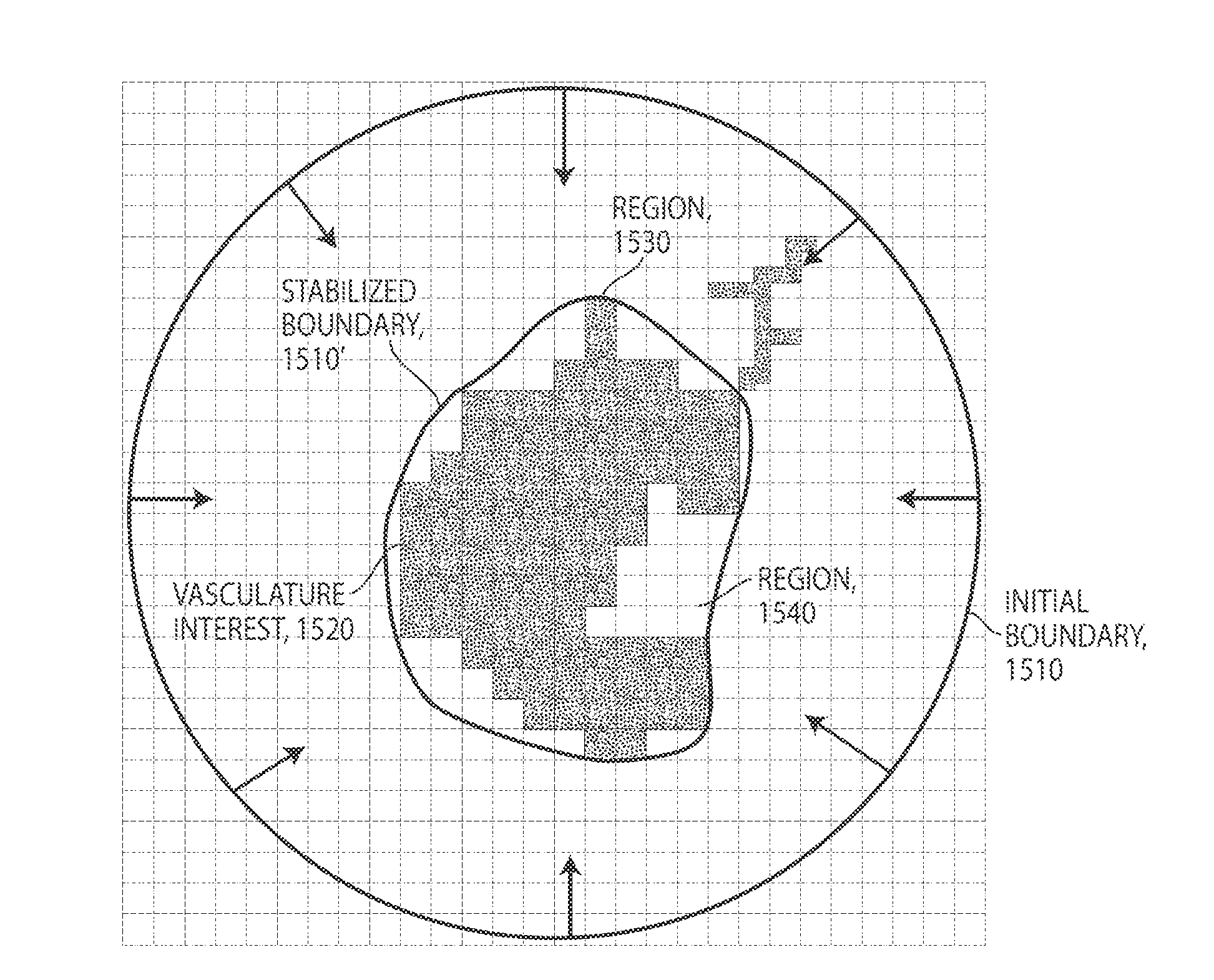
[0299]Some aspects relate to biomarkers identified in blood vessel structure. Some aspects of relate to analyzing vascular structure, for example, by assessing vascular biomarkers, using the Poker Chip representation (i.e., one exemplary type of 3D geometric representation of vasculature (see, for example, FIGS. 35-42 and 43-47). Some biomarkers provided by aspects of this invention relate to structural parameters of blood vessels (see, for example, the Figures). Some biomarkers provided by aspects of this invention are based on voxel (unit of volume) analysis of a given volume (see, for example, FIGS. 48-54). For example, a voxel may be associated with information related to vascular structure (e.g., vessel density), and voxel analysis of a given tissue volume may be used for continuous mapping of the vasculature in said tissue volume. In some embodiments, the boundary of a diseased tissue, for example a tumor, is determined, for example by determining a tumor wr...

example 3

Vasclular Biomarkers

[0300]Some aspects provide biomarkers of vasculature structure useful to identify and precisely locate abnormalities in vasculature, for example abnormalities associated with malignant tissue. Some non-limiting examples of vasculature structure biomarkers in accordance to this invention may be related to vascular organization, vascular density, and / or vascular anatomy. Some vascular biomarkers provided by this invention are related to micro-vasculature, for example to micro-vascular organization, micro-vascular density, and / or micro-vascular anatomy.

[0301]Some non-limiting examples of vascular organization biomarkers according to some aspects of this invention are vascular hierarchy (for example distribution of vascular hierarchy bins over a given tissue volume, frequency of a given vascular hierarchy bin within a given tissue volume), vascular branching (for example number of blood vessel branching points over a given blood vessel length, branching point density...


PUM


Abstract

According to some aspects, a method of identifying a boundary of a portion of a vasculature is provided, the vasculature comprising a geometric representation of a plurality of vessels. The method comprises logically dividing the geometric representation into a plurality of regions, determining at least one feature within each of the plurality of regions, and defining the boundary of the portion of the vasculature based, at least in part, on the at least one feature determined within each of the plurality of regions, wherein the boundary forms a volume defining a separation between inside and outside of the portion of the vasculature. According to some aspects, a method of performing vascular analysis using a geometric representation of a plurality of vessels of the vasculature is provided. The method comprises computing a boundary of a portion of the vasculature based on the geometric representation, logically dividing the geometric representation within the boundary into a plurality of regions, and analyzing at least one feature for each of the plurality of regions within the boundary.

Description

RELATED APPLICATIONS[0001]This application claims the benefit under 35 U.S.C. §119(e) from U.S. provisional application Ser. No. 61 / 209,386 entitled “Vascular Biomarkers for Diagnostic and Therapeutic Use” filed Mar. 6, 2009, the disclosure of which is incorporated herein by reference in its entirety.BACKGROUND OF THE INVENTION[0002]A wide range of imaging methods and devices are commonly used to evaluate different anatomical and physiological conditions in a variety of medical and research environments. Tools have been developed to image body structures based on different physical properties. For example, X-rays, CT scans, MRIs, PET scans, IR analyses and other technologies have been developed to obtain images of various body structures. These tools are routinely used for diagnostic, therapeutic, and research applications. Combinations of two or more different imaging techniques are sometimes used to provide complementary information about a patient.[0003]In conventional medical im...

Claims


Application Information

Patent Timeline
Patent Type & Authority Applications(United States)
IPC IPC(8): G06T7/00
CPCG06T7/0016G06T7/0081G06T7/0091G06T2207/30101G06T2207/20112G06T2207/20021G06T2207/20036G06T2207/10072G06T2207/30172A61B6/508G06T7/11G06T7/13G06T7/149G06T7/155
Inventor BRAUNER, RAUL A.KANG, KONGBINWU, YANCHUNCRUZ, JOAO
Owner BIO TREE SYST
Why Eureka
Try Eureka