
Adhesive-Mountable Head-Wearable EEG Apparatus

a technology of physiological monitoring and adhesive mounting, which is applied in the field of headwearable physiological monitoring devices, can solve the problems of easy dislocation, delicate wired sensors, and patients' dislike of wired sensors, and achieve the effects of improving the state of the art, reducing the degree of miniaturization, and adding comfort and user-friendliness

Inactive Publication Date: 2015-12-31
VASAPOLLO CURZIO
View PDF4 Cites 43 Cited by
  • Summary
  • Abstract
  • Description
  • Claims
  • Application Information

AI Technical Summary

Benefits of technology

The invention is an EEG apparatus that improves user-friendliness and comfort during sleep-staging. It achieves this through an adhesive electrode assembly with multiple electrodes that allow for easier use and reduced likelihood of disconnection. The electrodes support and mechanically mount a compact EEG device to the forehead, making a supporting headband unnecessary in most cases. The apparatus is multi-channel, low noise, wearable during sleep, and can be easily operated with no assistance. It also allows for easy acquisition of pulse oximetry in addition to EEG. Additionally, the enclosing shell has a chamfered edge to reduce lateral forces on the shell during sleep.

Problems solved by technology

Wired sensors are disliked by patients, and when they are used during sleep they disturb the process they are meant to monitor.
Further, wired sensors are delicate, and can become dislodged easily when the patient moves; their use normally requires trained staff.
It is perhaps due to these difficulties that sleep test devices designed specifically for home use, such as the Clevemed® SleepView™, Novasom® Accusom™, the Watermark® ARES™ and others, do not include EEG sensors.
Due to the high impedance of the conductive textile sensors used in the Zeo™, users reported inaccurate readings, high noise, and poor electrode performance.
Furthermore, users with long hair reported problems with the headband's stability and comfort.
None are suitable for wearing while the subject is sleeping due to their construction.
The Melon™ headband has only one channel, and the thickness of the device would make it difficult to wear during sleep.
The Muse™ hides the bulk of the device behind the ears, again making it unsuitable for wearing during sleep.

Method used


Image

  • Adhesive-Mountable Head-Wearable EEG Apparatus
  • Adhesive-Mountable Head-Wearable EEG Apparatus
  • Adhesive-Mountable Head-Wearable EEG Apparatus

Examples


Embodiment Construction

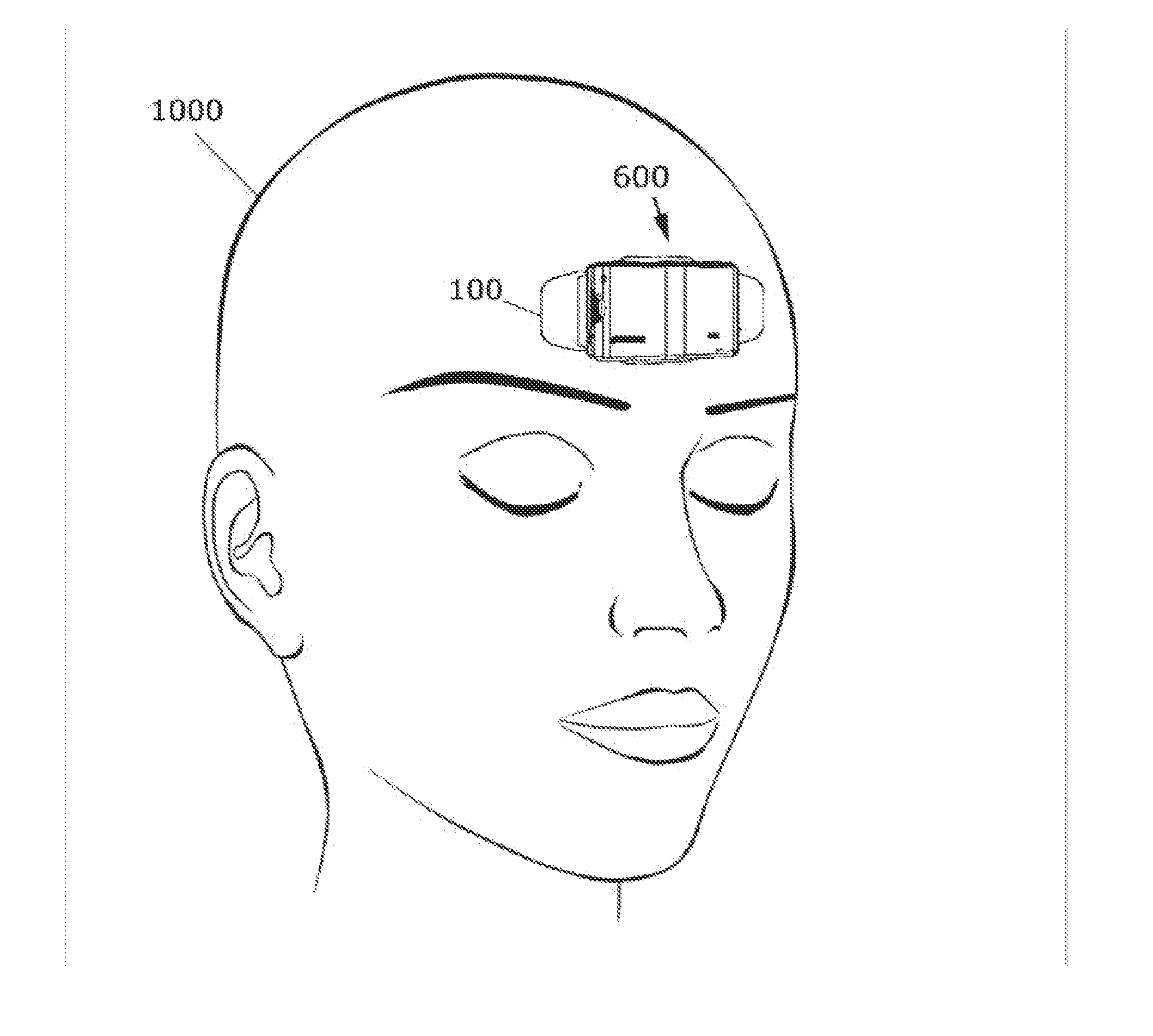
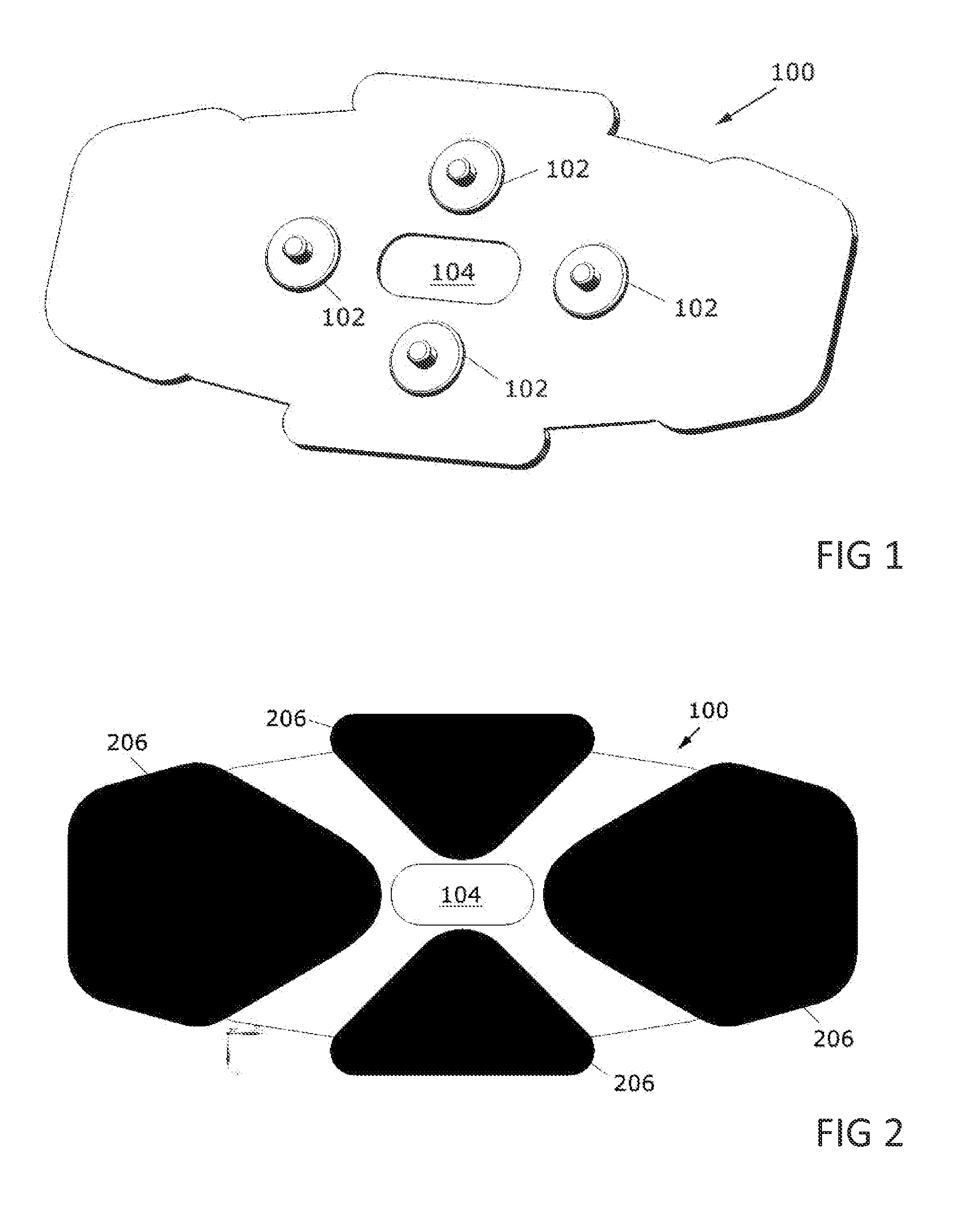
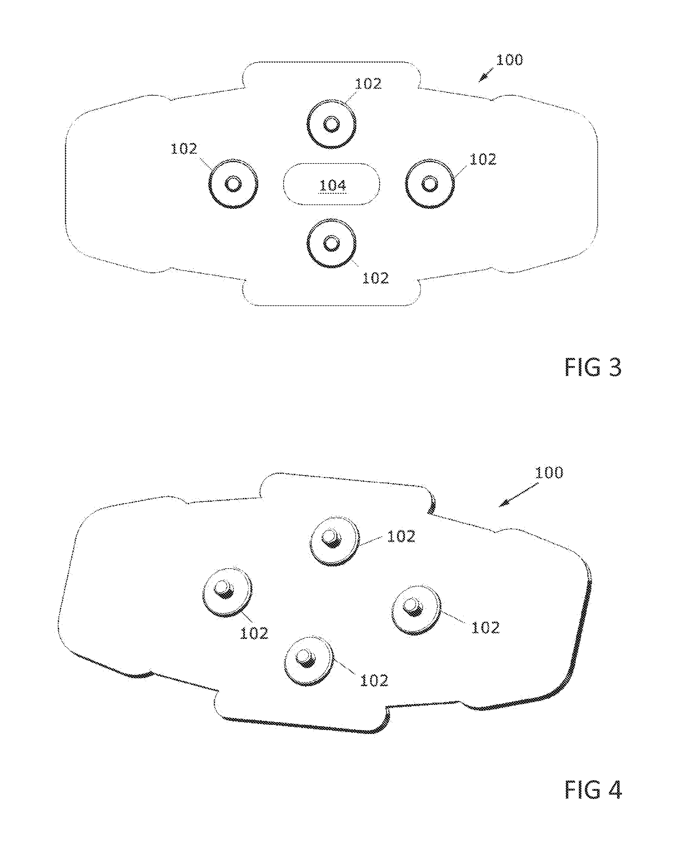
[0052]With reference to FIGS. 1-3, an adhesive electrode assembly 100 has four connection elements 102. A single adhesive electrode assembly 100 can be affixed to the forehead of a wearer easily and reliably even by inexperienced users, and reduces the possibility of individual electrodes becoming disconnected. The EEG signal is acquired from the wearer's forehead through the four gel electrodes 206. The electrical potential on the left and right gel electrodes 206 is measured against a reference electrode (either the central top or central bottom electrode 206). The remaining central top or bottom electrode 206 (the electrode not used as reference electrode) is an output from the EEG monitoring device, having “right leg drive” function to reduce common mode noise. Therefore the four electrodes 206 are used to acquire the left and right hemisphere frontal EEG signal (two channels of data), with low noise.

[0053]FIG. 4 shows an alternate embodiment of the adhesive electrode assembly o...


PUM


Abstract

An adhesive-mountable head-wearable EEG apparatus is disclosed. The apparatus includes an EEG sensor for acquiring an EEG signal of a wearer, a central processing unit for receiving the EEG signal, a small circuit board including the EEG sensor and the central processing unit, and a compact enclosing shell for enclosing the small circuit board, the EEG sensor, and the central processing unit. An adhesive electrode assembly attaches to the compact enclosing shell, or to the small circuit board within the enclosing shell, via snaps or magnets. The adhesive electrode assembly includes two or more gel electrodes for acquiring an EEG signal, and for adhering to the forehead so as to wearably support the EEG apparatus on the forehead. The compact enclosing shell includes chamfered edges, and is sized so as to reduce lateral forces on the compact shell that would tend to detach the EEG apparatus from the wearer's forehead.

Description

FIELD OF THE INVENTION[0001]This invention relates to head-wearable physiological monitoring devices, and particularly to such devices that include EEG monitoring.BACKGROUND OF THE INVENTION[0002]Physiological monitoring of a kind and accuracy once possible only in a clinical setting and with the aid of trained medical staff is becoming available as wearable consumer devices. This phenomenon promises to bring many benefits. First, lowered costs for patients. Second, the possibility to acquire physiological measurements over a longer time span, with benefits for both research and diagnosis. Third, the possibility of monitoring physiological parameters in a “real life” setting as opposed to an artificial laboratory setting. Fourth, the possibility to monitor physiological parameters with less discomfort, without disturbing the patient and compromising the data being acquired. Fifth, because of lowered costs and increased comfort, the benefits of physiological monitoring and early diag...

Claims


Application Information

Patent Timeline
Patent Type & Authority Applications(United States)
IPC IPC(8): A61B5/0478A61B5/00A61B5/1455
CPCA61B5/0478A61B5/14551A61B5/6814A61B5/6833A61B2562/06A61B5/291
Inventor VASAPOLLO, CURZIO
Owner VASAPOLLO CURZIO
Learn More