Method of Enhancing Delivery of Therapeutic Compounds to the Eye

a technology of therapeutic compounds and eye, which is applied in the direction of antibody medical ingredients, algae medical ingredients, peptide/protein ingredients, etc., can solve the problems of limited delivery effect of current methods, such as intravitreal injection or implanted drug delivery devices, and achieve the effects of improving or restoring light sensitivity, improving or restoring vision

Active Publication Date: 2016-02-11
WAYNE STATE UNIV
View PDF1 Cites 5 Cited by
  • Summary
  • Abstract
  • Description
  • Claims
  • Application Information

AI Technical Summary

Benefits of technology

[0009]In one aspect, the therapeutic agent is selected from a small molecule, a nucleic acid, an antibody, or a peptide. The nucleic acid is a nucleic acid expression vector (i.e., a viral vector), a plasmid, or an siRNA. For example, the viral vector is a AAV viral vector (i.e., recombinant AAV or rAAV) that encodes a transgene. Preferably, the transgene encodes a gene product that increases or restores light sensitivity, increases light detection, increases photosensitivity, increases visual evoked potential, or restores vision to the blind. More preferably, the transgene is an opsin gene. Examples of opsin genes include, but are not limited to, channelrhodopsins (i.e., channelrhodopsin-1, channelrhodopsin-2, Volvox carteri channelrhodopsins 1 or 2), melanopsin, pineal opsin, photopsins, halorhodopsin, bacteriorhodopsin, proteorhodopsin, or any functional variants or fragments thereof.
[0014]The present invention further provides a method of increasing or restoring light sensitivity in a subject comprising administering a plasmin or derivative thereof and a viral vector that encodes an op sin to the vitreous of the eye. The present invention also provides a method of improving or restoring vision in a subject comprising administering a plasmin or derivative thereof and a viral vector that encodes an opsin to the vitreous of the eye.

Problems solved by technology

Delivery of therapeutic compounds to specific ocular tissues affected by an ocular disease or disorder, such as the retina, is a challenge.
Current methods, such as intravitreal injection or implanted drug delivery devices, are still limited in the efficacy of delivery.
Specifically, the therapeutic agents are often localized only to the immediate areas surrounding the delivery site, and fail to permeate or diffuse beyond intervening ocular structures or throughout the targeted ocular tissue, thereby severely limiting the efficacy of such therapeutics.

Method used


Image

  • Method of Enhancing Delivery of Therapeutic Compounds to the Eye
  • Method of Enhancing Delivery of Therapeutic Compounds to the Eye
  • Method of Enhancing Delivery of Therapeutic Compounds to the Eye

Examples


example 1

Plasmin Increases Delivery of AAV2-Vector Encoding Chop2 to the Retina

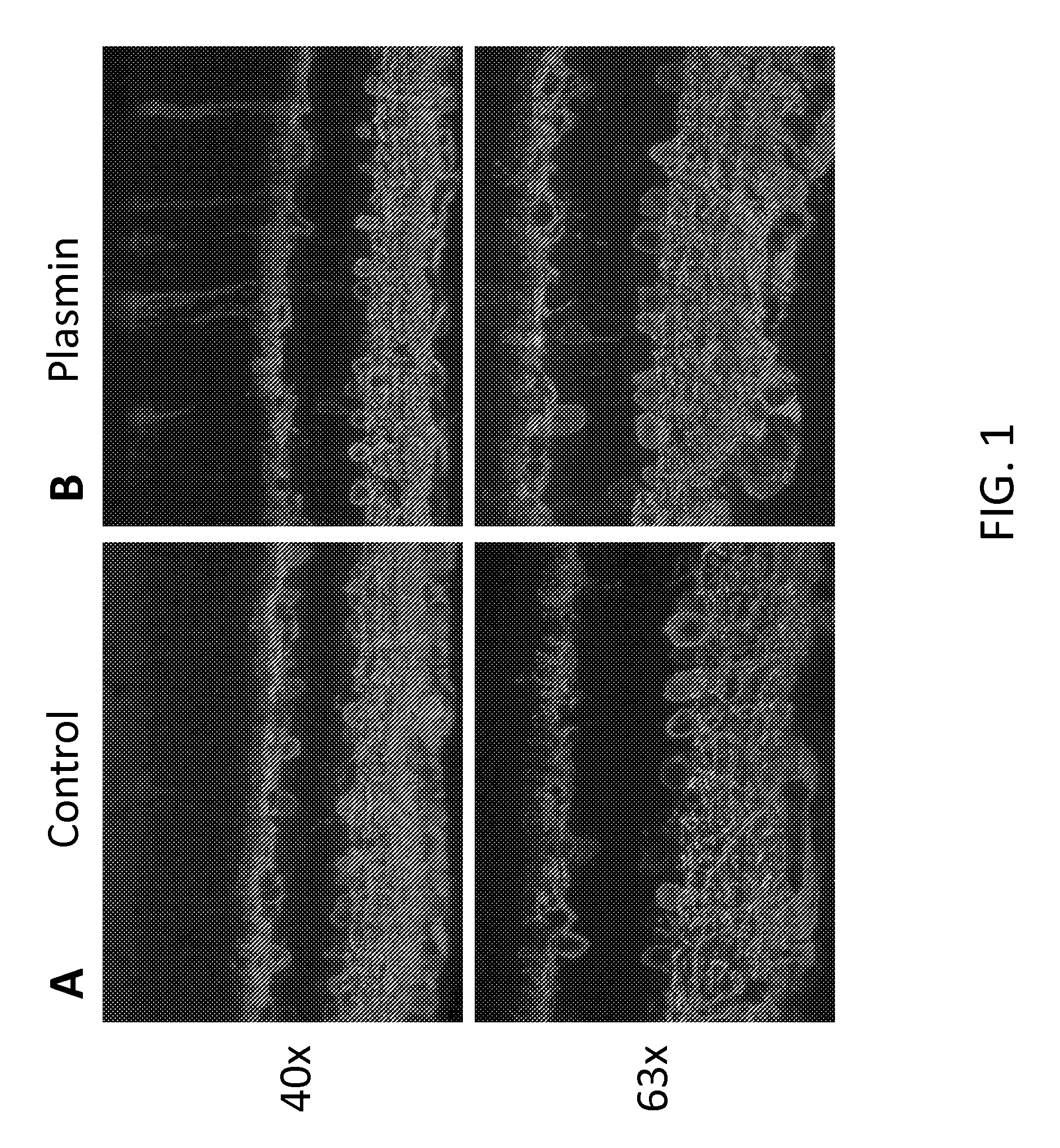
[0104]Delivery of a therapeutic viral construct encoding a functional GFP-channelopsin-2 protein to the retina was examined. Injection of 6×10¹² vg / ml of AAV2 vector AAV2 / 2-ChR2-GFP-WPRE-hGHpA in control (FIG. 1A) or co-injection with plasmin (FIG. 1B) was performed into the vitreous space of the eye of one month old C56BL / 6J. The concentration of plasmin injected with vector was 0.0251 U / eye. After one month, the mice retinas were isolated, and retinal vertical sections were prepared. The sections were immunostained and cells were counted. Immunofluorescence analysis of the sections showed that co-injection with plasmin increased the transduction efficiency of the therapeutic AAV2-ChR2-GFP vector, as evidenced by increased fluorescence in comparison to control (FIG. 1).

example 2

Plasmin Increases Transduction Efficiency in Retinal Ganglion Cells

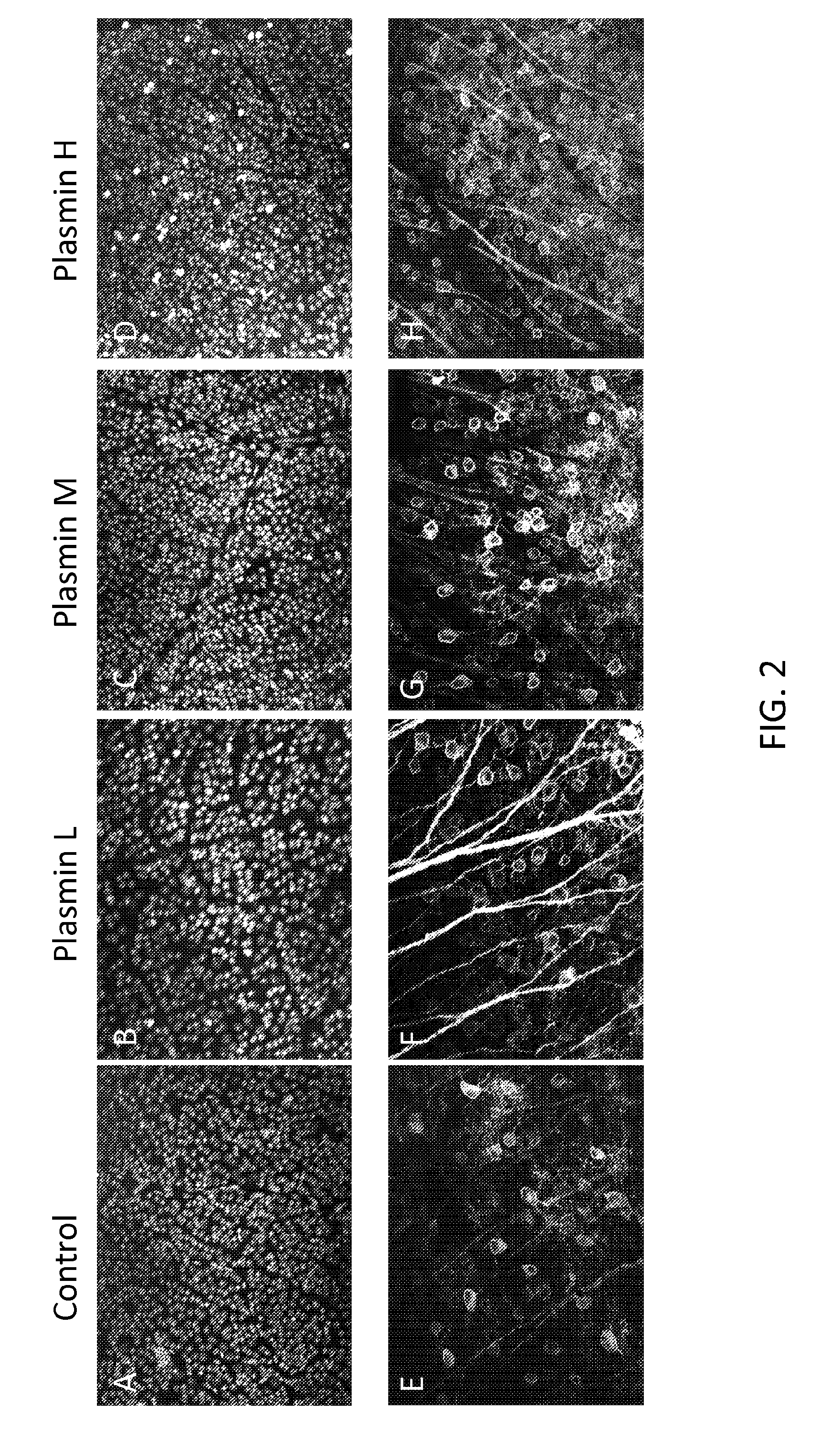
[0105]Using the same experimental set-up as in Example 1, a vector (2×10¹² vg / ml) encoding GFP was co-injected with either control or varying concentrations of plasmin: low (L=0.005 IU / eye), middle (M=0.0251 U / eye), and high (H=0.100 IU / eye). After 1 month, retinal whole mounts were prepared and immunostained. Representative images for each plasmin concentration and control are shown in FIG. 2. As shown, treatment with plasmin increases GFP expression.

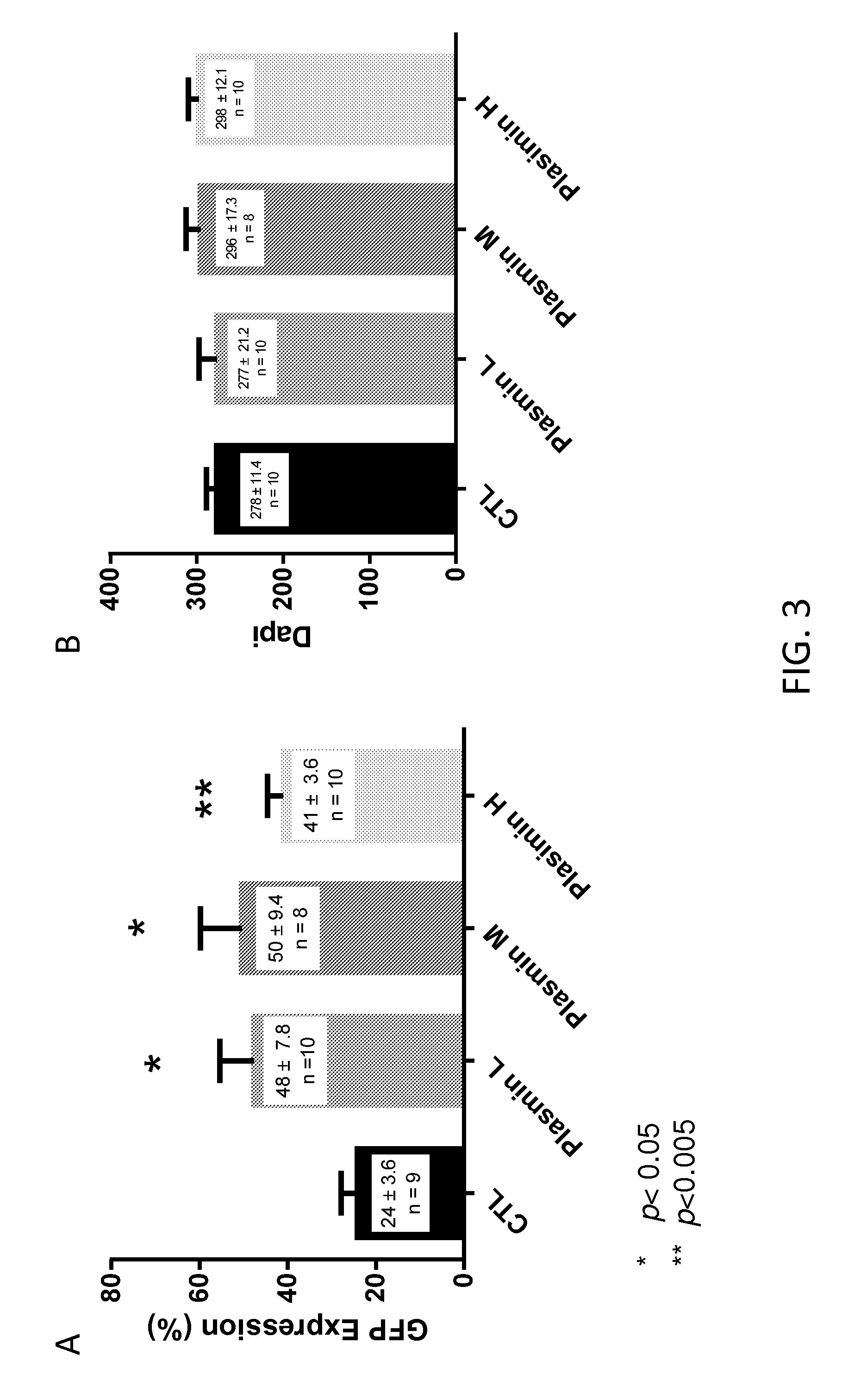
[0106]To further quantify these results, GFP-expressing retinal ganglion cells were counted from multiple unit areas of 223 μm×167 μm. The results are presented in FIG. 3A. As shown, treatment with low, middle and high doses of plasmin resulted in statistically significantly increased levels of GFP expression in retinal ganglion cells.

[0107]Neurotoxicity as a result of plasmin injection was also examined. The retinal whole mounts were stained with DAPI for cell-counting...

example 3

Plasmin Increases Transduction Efficiency in Retinal Bipolar Cells

[0108]Comparison of the transduction efficiency of a viral vector encoding mCherry fluorescent protein when co-injected with different concentrations of plasmin was assessed in vivo. Specifically, overall levels and the localization of mCherry expression throughout the retina were examined. An AAV2 vector with an Y444F capsid mutation carrying mCherry under control of an mGluR6 promoter were injected at a concentration of 2×10¹² vg / ml. The mGluR6 promoter directs expression of mCherry specifically to the retinal bipolar cells.

[0109]The AAV2 mCherry vector was co-injected with three doses of plasmin, high (H=0.100 IU / eye), middle (M=0.025 IU / eye), and low (L=0.005 IU / eye). After 1 month, the retinas were isolated and retinal whole-mounts were prepared. Transduction efficiency was evaluated by immunostaining of mCherry for immunofluorescence analysis and cell counting. Cells were counted from multiple unit areas of 223 ...


PUM

PropertyMeasurementUnit
volumeaaaaaaaaaa
nucleic acid expression vectoraaaaaaaaaa
photosensitivityaaaaaaaaaa

Abstract

The invention provides methods for enhancing the delivery of therapeutic compounds to the eye of a subject by administering plasmin or derivatives thereof and the therapeutic compounds to the eye.

Description

RELATED APPLICATIONS[0001]This application claims priority to, and benefit of, the U.S. Provisional Application No. 61 / 785,015 filed on Mar. 14, 2013; the contents of which are hereby incorporated by reference in its entirety.GOVERNMENT SUPPORT[0002]This invention was made with U.S. Government support under the National Institutes of Health / National Eye Institute grant NIH EY 17130. The Government has certain rights in the invention.FIELD OF THE INVENTION[0003]This invention relates generally to methods of enhancing the delivery of therapeutic compounds to the eye.BACKGROUND OF THE INVENTION[0004]The eye is a complex optical system that detects light, converts the light to a set of electrical signals, and transmits these signals to the brain, ultimately generating a representation of our world. Ocular diseases and disorders can cause diminished visual acuity, diminished light sensitivity, and blindness.[0005]Delivery of therapeutic compounds to specific ocular tissues affected by an...

Claims


Application Information

Patent Timeline
Patent Type & Authority Applications(United States)
IPC IPC(8): A61K9/00A61K36/05A61K38/16A61K38/48A61K48/00
CPCA61K9/0048A61K38/484A61K38/16C12Y304/21007A61K48/0008A61K48/0075A61K36/05A61K38/36A61K31/7088A61K31/713A61P27/00A61P27/02
Inventor PAN, ZHUO-HUAIVANOVA, ELENA
Owner WAYNE STATE UNIV
Why Eureka
Try Eureka