Homogenous thermal shift ligand binding assay

a technology of thermal shift and binding assay, which is applied in the direction of material analysis, instruments, biological material analysis, etc., can solve the problems of lack of solubility and accessibility of labeling peptides, and achieve the effect of high statistical significan

Active Publication Date: 2016-02-25
DISCOVERYX CORP
View PDF1 Cites 11 Cited by
  • Summary
  • Abstract
  • Description
  • Claims
  • Application Information

AI Technical Summary

Benefits of technology

[0133]The present experiment was done to study the importance of centrifugation to separate precipitated fusion protein in a composition also containing soluble, bound fusion protein, and how it affects the final readout.
[0134]To carry out the experiment, a fusion protein was constructed comprising NFκB-BRD4(1)-Prolabel (ED). The fusion protein was then incubated with a ligand and lysis was done using FLASH lysis and in PBS. The samples were then kept at room temperature or exposed to 45° C. elevated temperature. Each sample was divided into 2 wherein one part was centrifuged and other part of the sample was not centrifuged.
[0135]As can be seen from FIG. 6, there is little difference in the assay readout whether the sample was centrifuged to separate the precipitate or not. Therefore, the experiment shows that centrifugation is not required in the present assay and the ligand binding can be assayed using EFC in the crude cell extract.
[0136]The present experiment was done to determine the dose-dependence of ligand protein binding following the ligand binding assay as described in the present application.
[0137]To carry out

Problems solved by technology

The method will cause a lack of solubility and accessibility of the labeling peptide attached to the target macrom

Method used


Image

  • Homogenous thermal shift ligand binding assay
  • Homogenous thermal shift ligand binding assay
  • Homogenous thermal shift ligand binding assay

Examples


example 1

Measuring Binding of BRD4(1) ED Fusion Protein with a Known Ligand (JQ1)

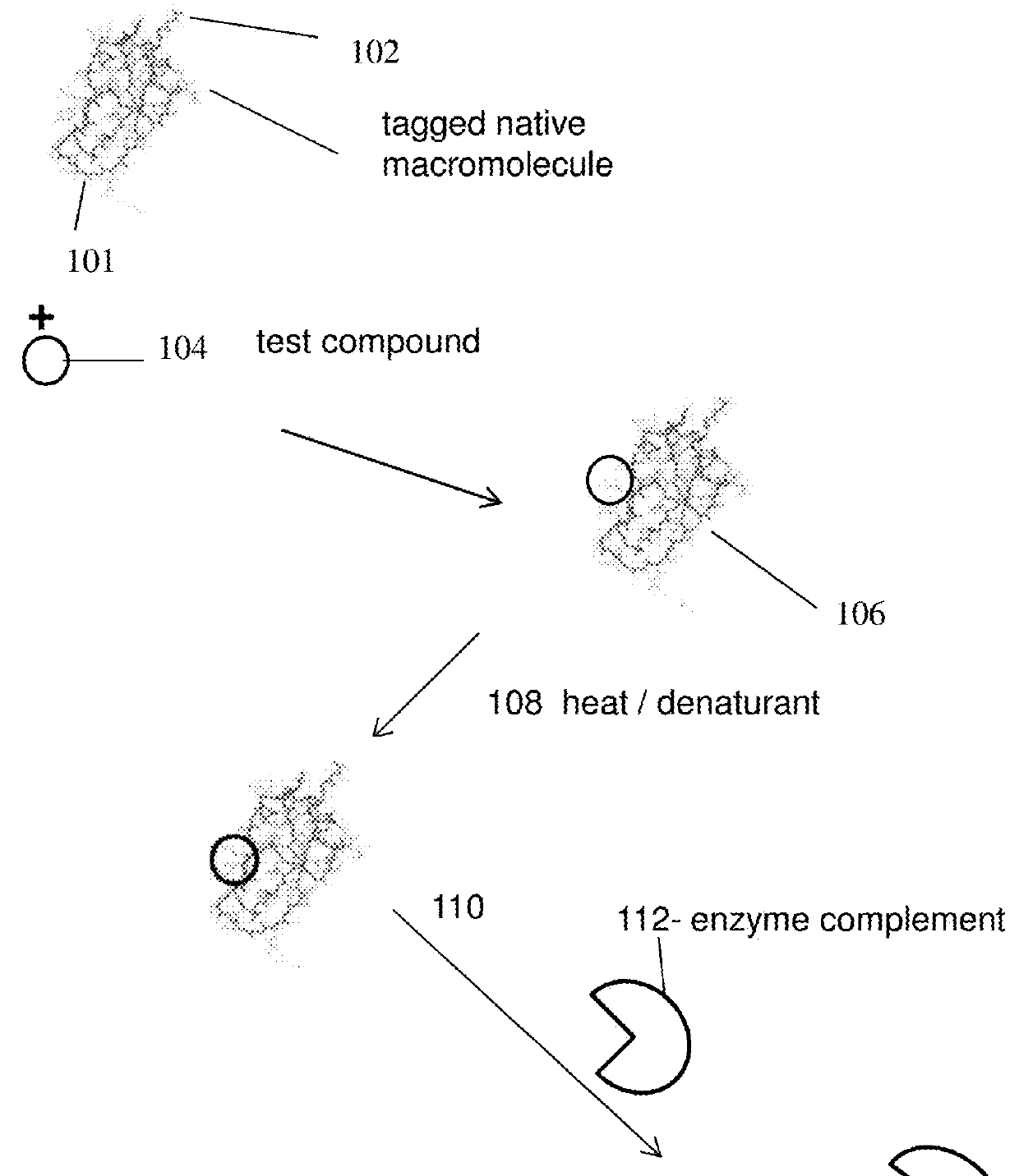
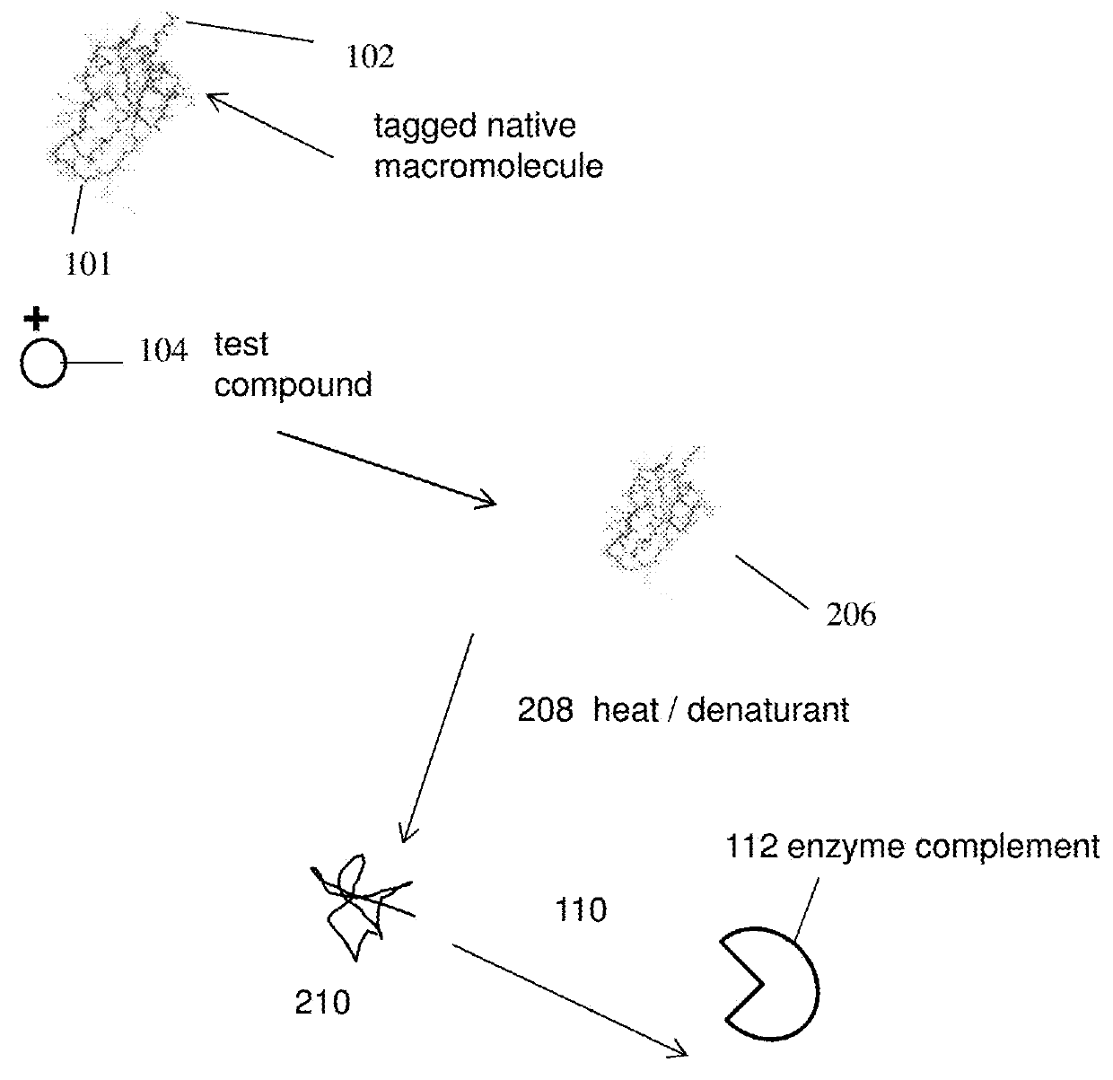
[0127]The present experiment was done to study binding of a ligand to BRD4(1) fusion protein under heat stress and if the ligand binding protects the fusion protein from denaturation.

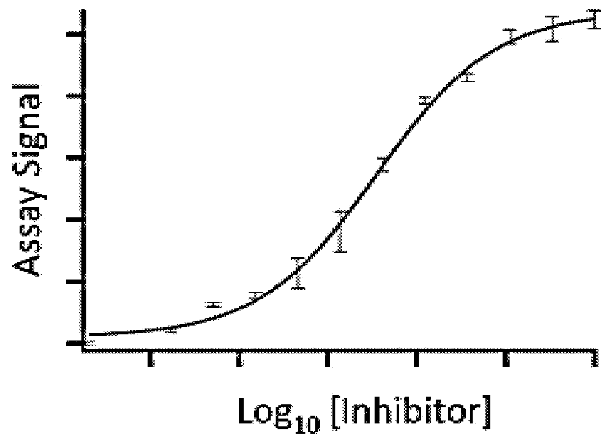
[0128]To carry out the assay, a fusion protein [BRD4(1)-ED], which also contains an NFκB DNA binding domain at the N-terminus, was constructed with BRD4(1) and a Prolabel™ ED fragment. The fusion protein in crude cell extract was incubated with a known potent inhibitor (JQ1) in a concentration ranging from 0-100 3M. The incubation was for 1 hour at room temperature. The samples were then heated to 45° C. for 30 s. EA was subsequently added into the samples (along with luminescent substrate for beta-galactosidase) and complementation was measured by measuring luminescence. As shown in FIG. 3, binding of the inhibitor to BRD4(1) fusion protein and binding could rescue the fusion protein from denaturation in a dose-dependent manner. Ex...

example 2

Measuring Thermal Denaturation of BRD4(1) in the Presence and Absence of JQ1

[0129]The present experiment was done to study denaturation / precipitation of BRD4(1) in the presence and absence of an inhibitor. The inhibitor as used in the present experiment is JQ1.

[0130]To carry out the assays, two separate samples were prepared wherein one sample comprises BRD4(1)(fusion) and no inhibitor whereas another sample comprises BRD4(1)(fusion) and an inhibitor. The inhibitor as used in the present experiment is JQ1 and was used at a concentration of 10 3M. Following incubation, samples were exposed to increased temperatures (25° C., 45° C., 50° C. and 55° C. respectively). The samples were then centrifuged to separate the precipitate. As can be seen from FIG. 4, JQ1 bound to the fusion protects BRD4(1) from precipitation as compared to BRD4(1) with no inhibitor binding.

example 3

Measuring Precipitation of BRD4(1) Fusion Protein in the Presence of JQ1

[0131]The present experiment was done to study how binding of JQ1 protects BRD4(1) fusion protein from temperature induced precipitation.

[0132]To carry out the experiment, a fusion protein was constructed comprising NFκB-BRD4(1)-ED. The fusion protein from cell lysate was incubated with JQ1 (10 μM). The samples were then exposed to several temperatures (25° C., 45° C., 50° C. and 55° C. respectively). As can be seen from FIG. 5, fold soluble BRD4(1) was maximally rescued at 45° C., and proteins slowly started precipitating at higher temperatures.


PUM


Abstract

Disclosed are methods for detecting and quantitatively measuring a binding property of a compound to a target macromolecule, wherein the target macromolecule is subject to denaturation and is linked to a labeling peptide, such as a short enzyme fragment. The method uses a fluid mixture comprising (i) a chimeric molecule comprising a target macromolecule linked to the labeling peptide and (ii) a compound being measured for binding to the target macromolecule, wherein said target macromolecule is subject to denaturation. After allowing for binding of the compound (e.g. a small molecule inhibitor of the target macromolecule), one detects a signal from the labeling peptide, such as by enzyme fragment complementation. This signal indicates a differential between denatured and non-denatured target macromolecules and thereby indicates a differential between target macromolecules not bound to the compound and target macromolecules bound to the compound, respectively.

Description

CROSS-REFERENCE TO RELATED APPLICATIONS[0001]This application claims priority from U.S. Provisional Patent Application No. 62 / 087,671, filed Dec. 4, 2014, and U.S. Provisional Patent Application No. 62 / 040,294, filed Aug. 21, 2014, both of which are hereby incorporated by reference in their entirety.STATEMENT OF GOVERNMENTAL SUPPORT[0002]None.REFERENCE TO SEQUENCE LISTING, COMPUTER PROGRAM, OR COMPACT DISK[0003]The instant application contains a Sequence Listing which has been submitted as an ASCII text file and is hereby incorporated by reference in its entirety. This text file was created on Aug. 18, 2015, is named “3817—54—1_seq_list.txt” and is 2,524 bytes in size.BACKGROUND OF THE INVENTION[0004]1. Field of the Invention[0005]The present invention relates generally to methods of monitoring binding of a compound to a target macromolecule by increasing the thermal stability of the target macromolecule. The present invention also relates to a novel method of use of enzyme fragment...

Claims


Application Information

Patent Timeline
IPC IPC(8): G01N33/573G01N33/68
CPCG01N33/573G01N33/6875G01N2500/04G01N2333/91017G01N2333/91057G01N2333/912G01N2333/918G01N33/542G01N2333/938G01N33/581H05K999/99
Inventor TREIBER, DANIEL K.MENICHELLI, ELENA
Owner DISCOVERYX CORP
Why Eureka
Try Eureka