Tube-detector alignment using light projections

a technology of light projection and x-ray tube, which is applied in the field of alignment of x-ray tube and x-ray detector, can solve the problems that the usability of such arrangement solutions in clinical practice is often very limited, and achieve the effect of simplifying the process of aligning an x-ray tub

Active Publication Date: 2016-03-17
KONINKLJIJKE PHILIPS NV
View PDF3 Cites 11 Cited by
  • Summary
  • Abstract
  • Description
  • Claims
  • Application Information

AI Technical Summary

Benefits of technology

[0051]It can be seen as an idea of the invention, to simplify the process of the alignment of an X-ray tube and a detector during a mobile X-ray imaging application by using intuitive light patterns which can be brought into alignment by medical staff, in order to achieve a correct arrangement of an X-ray tube and a detector relative to each other.

Problems solved by technology

However, the practical usability of such arrangement solutions in clinical practice can often be very limited.

Method used


Image

  • Tube-detector alignment using light projections
  • Tube-detector alignment using light projections
  • Tube-detector alignment using light projections

Examples


Embodiment Construction

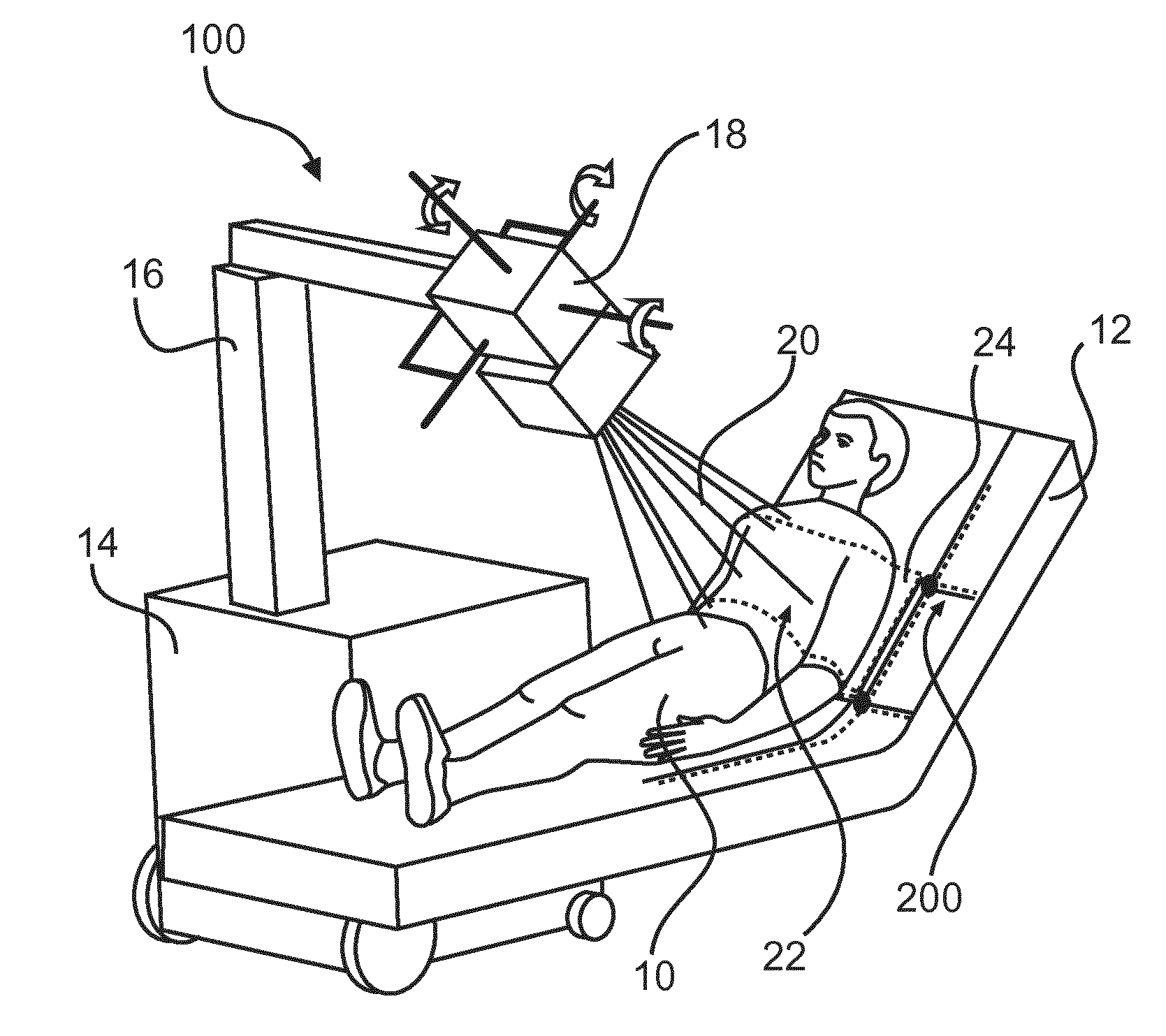
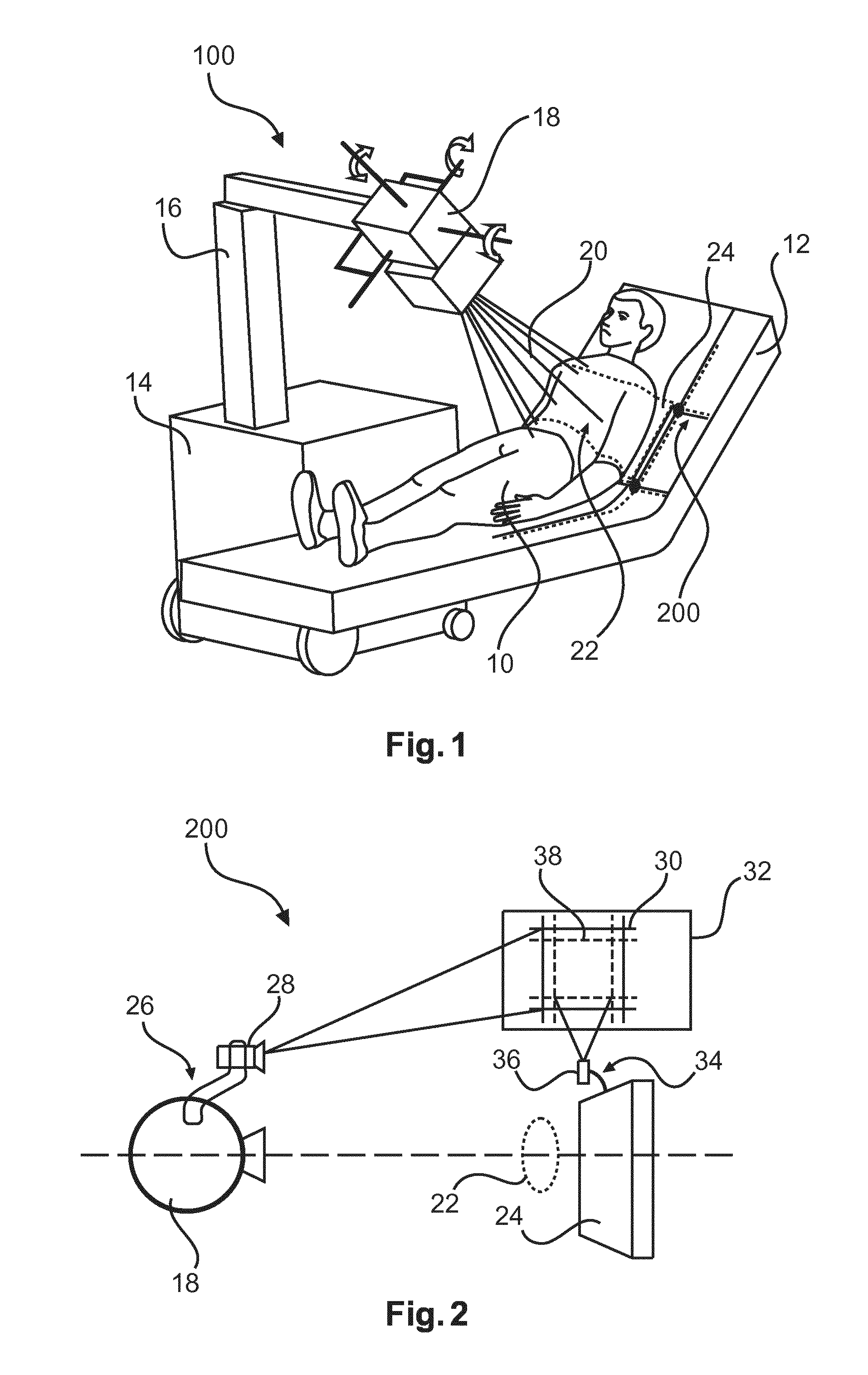
[0062]In FIG. 1, a medical X-ray imaging system 100 is shown in a bedside medical imaging situation. In the shown example, medical image information is acquired of an object 10 positioned on a patient support 12, wherein the patient support 12 also serves as projection surface. The imaging system 100 comprises a movable base unit 14, a support arm 16, and a spatially adjustable X-ray tube 18. The X-ray tube 18 generates and emits X-ray 20 towards the object 10 and irradiates a region of interest 22. A detector 24 is positioned between the patient support and the region of interest 22. The detector 24 can be, for example, a portable detector panel, which can have a cable or wireless data connection and / or a power supply connection with the base unit 14. In preparation for a medical imaging, the portable detector is manually positioned behind the region of interest 22. For this purpose, it may become necessary for the patient to raise or lift parts of his body, for instance, the upper...


PUM


Abstract

The present invention relates to acquisition of medical image information of an object. In order to provide a user-friendly alignment of X-ray tube (18) and a detector (24), optionally combined with an anti-scatter grid, an alignment arrangement (200) is proposed, which comprises a tube attachment (26) with a first light projection device (28) and a detector attachment (34) with a second light projection device (36). The first and second light projection devices each generate a light pattern (30, 38) on a projection surface (32). The tube attachment (26) and the detector attachment (34) can be brought into a correct spatial arrangement relative to each other by bringing the first light pattern in a predetermined spatial relation with the second light pattern (38) on the projection surface.

Description

FIELD OF THE INVENTION[0001]The present invention relates to medical image information acquisition of an object. In particular, the present invention relates to an alignment of an X-ray tube and an X-ray detector, in particular for mobile X-ray imaging applications. The invention further relates to a method for aligning an X-ray tube relative to a detector.BACKGROUND OF THE INVENTION[0002]In clinical practice, mobile X-ray imaging systems are used in cases, where patients are unable to leave the patient's bed and where it may become necessary to conduct medical imaging diagnostics in bedside situations. Such mobile X-ray imaging units can comprise a movable X-ray base unit, which may further be equipped with further functions and applications, such as image processing or control functions. For example, a movable base unit can further comprise a support arm, where an X-ray tube is mounted to. A separate detector or detector panel receives the radiation, which has irradiated a, for in...

Claims


Application Information

Patent Timeline
Patent Type & Authority Applications(United States)
IPC IPC(8): A61B6/00A61B6/08
CPCA61B6/587A61B6/4405A61B6/08A61B6/0492A61B6/461A61B6/547A61B6/585A61N5/1048A61N2005/105G02B7/005G02B27/64H01J37/22H01J2237/1501H01J2237/20292H01J2237/24528H01J2237/2482H04B10/11
Inventor MANKE, DIRKKURZE, CHRISTOPHVAN DE VEN, RICHARD JOHANNUS MARIA
Owner KONINKLJIJKE PHILIPS NV
Why Eureka
Try Eureka