

Apparatus and methods for identifying and evaluating bright spot indications observed through optical coherence tomography
a technology of optical coherence tomography and optical coherence tomography, which is applied in the field of apparatus and methods for identifying and evaluating bright spot indications observed through optical coherence tomography, can solve the problems that the risk of plaque rupture cannot be easily assessed by only ivoct images, and the combination of fiber-based oct-tpl system has not been previously realized
- Summary
- Abstract
- Description
- Claims
- Application Information
AI Technical Summary
Benefits of technology
Problems solved by technology
Method used
Image

Examples
example 1
IVOCT Bright Spot Quantification and Analysis
[0076]It is hypothesized that bright spots in intravascular optical coherence tomography (IVOCT) images may originate by co-localization of plaque materials of differing indices of refraction (IR). To quantitatively identify bright spots, an algorithm according to the present disclosure was developed that accounts for factors including tissue depth, distance from light source, and signal-to-noise ratio. This algorithm was used to perform a bright spot analysis of IVOCT images, and to compare these results with histologic examination of matching tissue sections
[0077]Although bright spots are thought to represent macrophages in IVOCT images, studies of alternative etiologies have not been reported.
[0078]Fresh human coronary arteries (n=14 from 10 hearts) were imaged with IVOCT in a mock catheterization laboratory then processed for histologic analysis. The quantitative bright spot algorithm was applied to all images.
[0079]Results are report...
example 2
A Quantitative Assessment Using Bright Spot Algorithm
[0133]Referring now to FIGS. 26 and 27, a study was performed to evaluate the statin efficacy on the change of plaque complexity using an OCT bright spot algorithm according to the present disclosure. Recent studies using OCT have reported that statin therapy increases the thickness of fibrous caps in human coronary plaques, but the efficacy on the stabilization of plaque complexity is unknown.
[0134]Fifty-nine lipid-rich coronary plaques in non-culprit lesions from forty patients were randomized to receive statins (specifically, 60 mg atorvastatin (AT60), 10 mg rosuvastatin (RT10), or 20 mg atorvastatin (AT20)). The images and data shown in FIGS. 26 and 27 were obtained at an initial baseline, six month and twelve month time periods. An OCT algorithm was applied that is able to identify bright spots representing a variety of plaque components, including for example, macrophages. The bright spot density was measured within superfic...
example 3
Bright Spot Representative Images and Spatial Distribution
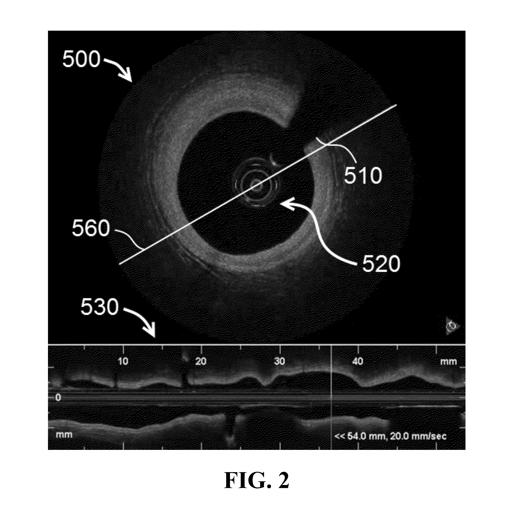
[0138]Referring now to FIG. 28, representative images and spatial distribution of bright spots are shown. Sections (a) and (b) of FIG. 28 provide representative images of specimens with acute coronary syndrome. Section (a) is a raw cross sectional OCT image, while section (b) provides a processed image using a bright spot algorithm as disclosed herein to a depth of 250 μm. The lower panel of section (b) represents the magnified image of the inset box shown in the upper panel of section (b). As shown in the figures, bright spot accumulation can be seen around the disrupted fibrous cap. A representative distribution of bright spot density within the longitudinal region of interest in a case of acute coronary syndrome is shown in section (c) of FIG. 28, with the X-axis representing the distal to proximal frame numbers.
[0139]Sections (d) and (e) of FIG. 28 provide representative images of specimens with stable angina pectoris. Ag...
PUM
 Abstract
Description
Claims
Application Information