
System and Method for Measurement of Myocardial Mechanical Function

Active Publication Date: 2016-04-07
TECHNION RES & DEV FOUND LTD
View PDF10 Cites 36 Cited by
  • Summary
  • Abstract
  • Description
  • Claims
  • Application Information

AI Technical Summary

Benefits of technology

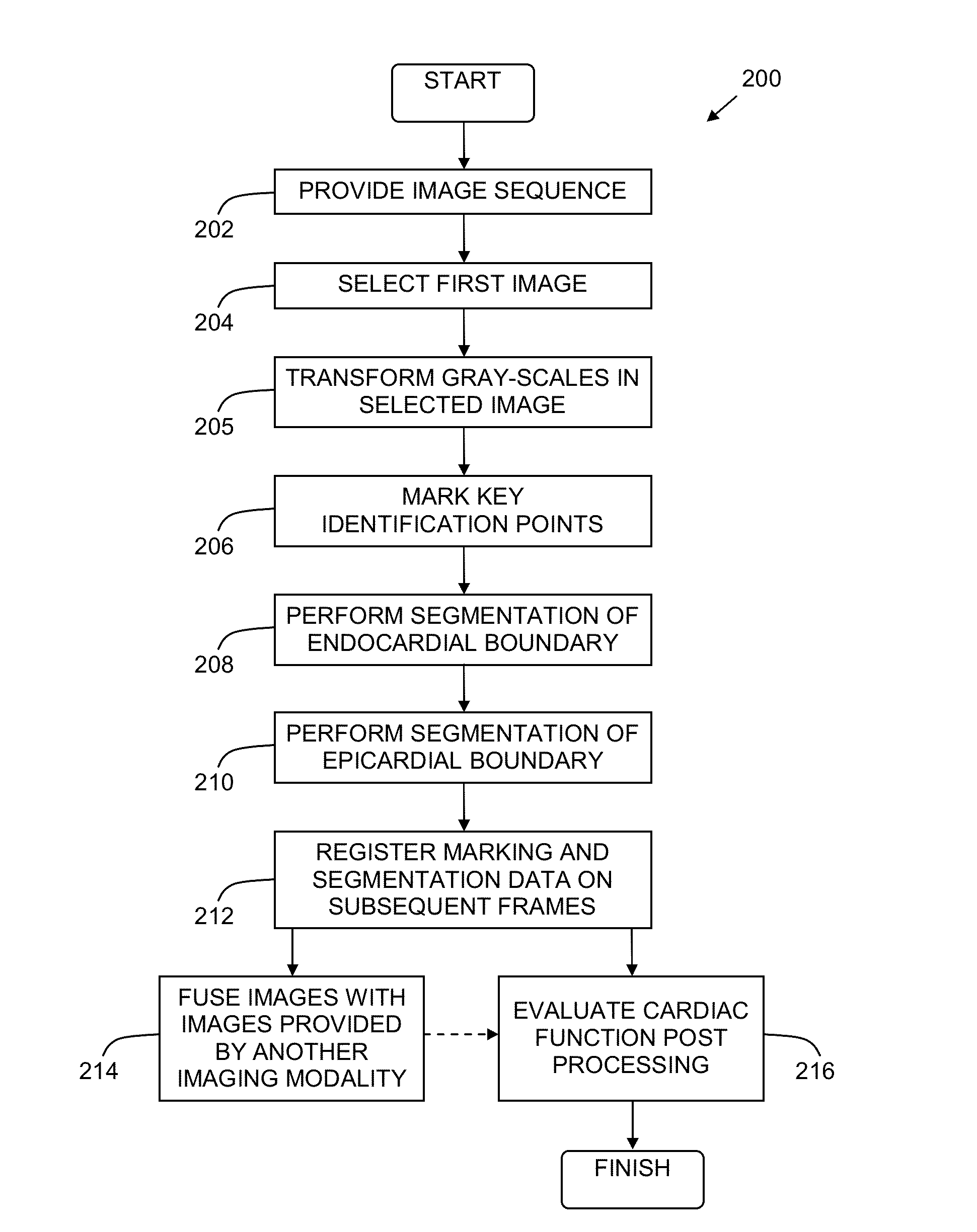
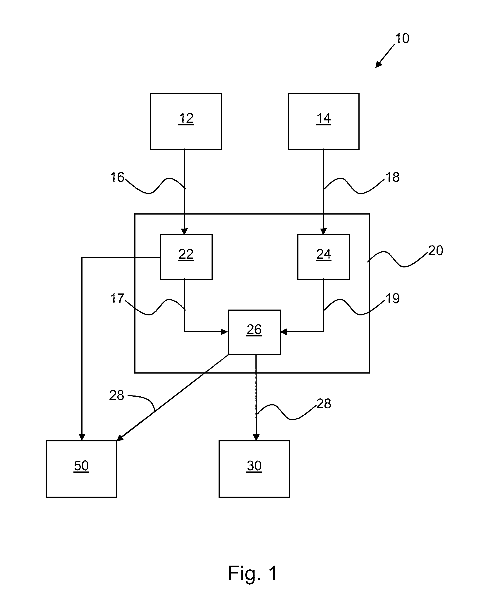
The present invention provides a method and system for evaluating cardiac function using image sequences obtained from different modalities, such as cine MRI, ultrasound, and cardiac CT. The method involves segmenting the image sequences, fusing them together, and analyzing the fused image to evaluate cardiac function. The system includes an imaging system and a processor for processing and fusing the image sequences. The technical effects of the invention include improved accuracy and reliability in evaluating cardiac function and providing more effective treatment based on the evaluation.

Problems solved by technology

Tagged MRI is, however, not the modality of choice for assessing myocardial mechanical function due to poor resolution.
Furthermore, once a pacemaker has been implanted further MRI studies (e.g. for following up and future adjustments) are precluded.
To date, the tools used for cardiac imaging and diagnostics are limited to post-processing workstations, cardiovascular workstations with one specific modality (such as ultrasound, for example), or CV PACS system, which provides the means to store, access, view, interact and report on the results of CV imaging procedures.
Typically, the PACS systems has ‘multi-modality’ capability but limited ‘post-processing’ functionality.
The existing tools today do not provide segmentation solutions for “long-axis” MRI images (only for “short-axis” (closed forms) but with limited accuracy).
None of the existing tools provide for automatic processing of left ventricular segmentation, and automatic calculations of clinical indices (LV volume, stroke volume, etc.).
None of the existing tools provide for data for longitudinal deformation in MRI “long-axis” images, and thus no ability to calculate strains.
Moreover, none of the existing tools allow for fusion of LE-MRI with c-MRI or for fusion of echocardiography and c-MRI or other imaging modalities with one another.
Tagging can be used to measure myocardial strain, but quantitative analysis of tagged images is not straightforward.
Some disadvantages of tagging are that it requires a specific and unique acquisition protocol, it usually suffers from progressive deterioration of the tag signal during the cardiac cycle, and it requires long breath-hold acquisition and time-consuming post-processing.
One drawback common to all of these approaches, among others, is that they require acquisition of a specialized image dataset at the time of the examination, typically in addition to the standard cine CMR of the ventricles.
Thus, they require a priori planning, increase the duration of the examination, and cannot be applied retrospectively to existing CMR datasets.

Method used


Image

  • System and Method for Measurement of Myocardial Mechanical Function
  • System and Method for Measurement of Myocardial Mechanical Function
  • System and Method for Measurement of Myocardial Mechanical Function

Examples


example 1

Automatic Segmentation of Cardiac MRI Cines for Long Axis Views

[0074]Validation of a method of segmentation of the endocardium and epicardium in 2CH and 4CH views was performed. The method was validated on a clinical dataset by comparing its results to manual segmentation performed by experienced observers, which serves a gold-standard.

[0075]Method: Cardiac MRI Datasets: Data were obtained at two orthogonal cross-sections per phase, 2CH and 4CH. Each data sequence was acquired as 2D+time apical cine slice using ECG gated cine cMRI. The data consisted of clinical exams of 126 patients with suspected cardiac disease. The datasets were obtained from two sources: 26 patients were obtained from the Oslo University Hospital, Rikshospitalet, Oslo, Norway and dataset of 100 patients were obtained from the cohort. These data were randomly selected from the Cardiac Atlas Project (CAP) database. All images were acquired using the steady state free precession protocol (SSFP) and 1.5 Tesla MRI s...

example 2

Myocardial Strain Assessment by Cine Cardiac Magnetic Resonance Imaging Using Non-Rigid Registration

[0102]Validation of a Tool for assessment of longitudinal strain in standard CMR sequences was performed. The method was validated with TMRI and compared to speckle tracking echocardiography (STE) and to the extent of scar tissue.

[0103]Methods: A total of 67 individuals were included in this study. The validation group consisted of 15 patients with previously diagnosed myocardial infarction (mean age 69±12 years, 14 males) and 15 healthy volunteers without clinical history of cardiac disease (mean age 27±5 years, 12 males). All these subjects underwent CMR and TMRI for reference strain analysis (two sources: Cardiac Atlas Project, STACOM 2011 challenge and The University of Queensaland, Brinsbane, Australia). The second group was used for the comparison stage of the study. This group included 37 patients (mean age 67±14 years, 28 males), with previously diagnosed myocardial infarction...


PUM


Abstract

There is provided a system and method for evaluation of cardiac images, wherein enhanced evaluation of myocardial mechanical function is possible. The system and methods include methods for segmentation of cardiac images obtained via cMRI or other imaging modalities, wherein the segmentation allows for fusion of these images with images obtained from a different modality such as echocardiogram and / or LE-MRI. The fused images may then be used to provide a diagnosis or a recommendation for a procedure, such as implantation of a cardiac pacemaker. Moreover, follow-up evaluation may be done using only one imaging modality, such as echocardiogram, for example. The system and methods disclosed herein further provide additional post-processing, such as computation of mid-myocardial strain, which can further be useful in diagnosis and planning.

Description

FIELD OF THE INVENTION[0001]The present invention is directed to a system and method for providing measurement of myocardial mechanical function for cardiac diagnostics.[0002]More specifically, the present invention is directed to systems and methods for c-MRI myocardial wall segmentation, strain measurements and fusion with other imaging modalities such as LE-MRI and ultrasound for enhanced measurement of myocardial mechanical function.BACKGROUND[0003]An increasing number of cardiac patients undergo diagnostic procedures using different modalities, e.g. Ultrasound (US), Magnetic Resonance Imaging (MRI), Echocardiography (ECG), Electrophysiology (EP), etc. The diagnostic information provided by these procedures is often used to select patients for Implantable Cardiac Defibrillators (ICD).[0004]Identification of candidates for ICD has traditionally been based on the electrophysiologic properties of noninfarcted myocardial tissue and the factors that trigger the induction of lethal ar...

Claims


Application Information

Patent Timeline
IPC IPC(8): G06T7/00G06T3/00G06K9/46G06T7/20G06K9/62G06T11/60G06V10/46
CPCG06T7/0012G06K9/6201G06T11/60G06T7/0085G06T2207/30048G06T7/0028G06T7/20G06T3/0093G06K9/4604G06T2207/10076G06T2207/10081G06T2207/20221G06T7/38G06V10/471G06V10/46G06V2201/031G06F18/22G06T3/18
Inventor TSADOK, YOSSIADAM, DAN
Owner TECHNION RES & DEV FOUND LTD
Learn More