Image forming cytometer
a cytometer and image technology, applied in the field of image cytometry analysis, can solve the problems of difficult microscopy, poor contrast, and microscopy that requires specialised optical components, and achieves the effect of simple, effective and reliabl
- Summary
- Abstract
- Description
- Claims
- Application Information
AI Technical Summary
Benefits of technology
Problems solved by technology
Method used
Image
Examples
example 1
Image Cytometer
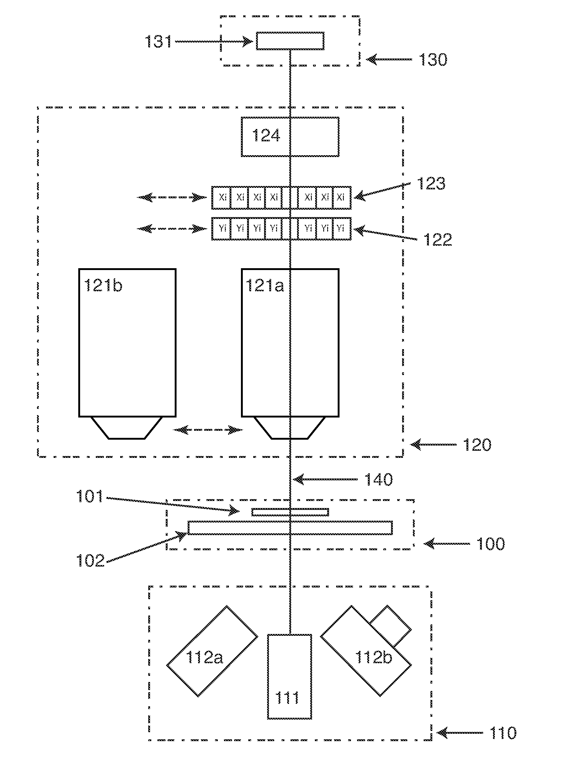
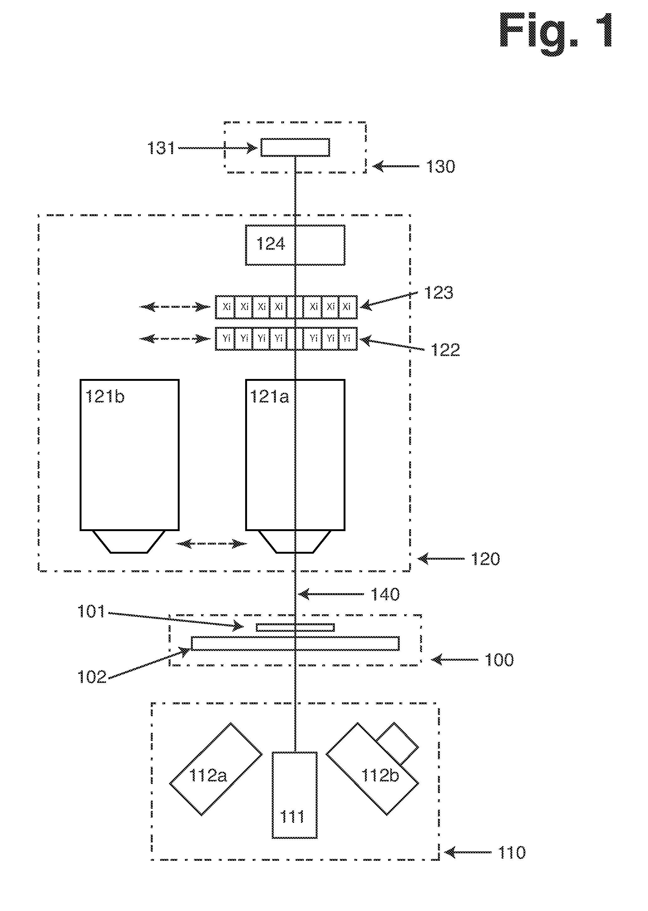
[0113]FIG. 1 illustrates possible configuration of an Image Cytometer including several preferred embodiments of the present invention. The illustration outlines 4 main groups of components, the sample holder (100), illumination means (110), imaging means (120) and detection means (130). Finally it illustrates the main optical axis of the Image Cytometer (140), along which majority of the optical components are arranged.
[0114]The sample holder can be moved along the optical axis relative to the imaging means, in order to assure that the sample is in focus alignment with the imaging means. The sample compartment (101) is placed on the sample stage (102) in the optical path. The sample compartment is typically attached to the sample stage but it can be released there from such that it can be removed from the Image Cytometer and replaced again through either manual or automated process. The sample stage can move in two directions perpendicular to the optical axis. This a...
example 2
Properties of Low Wavelength Microscopy
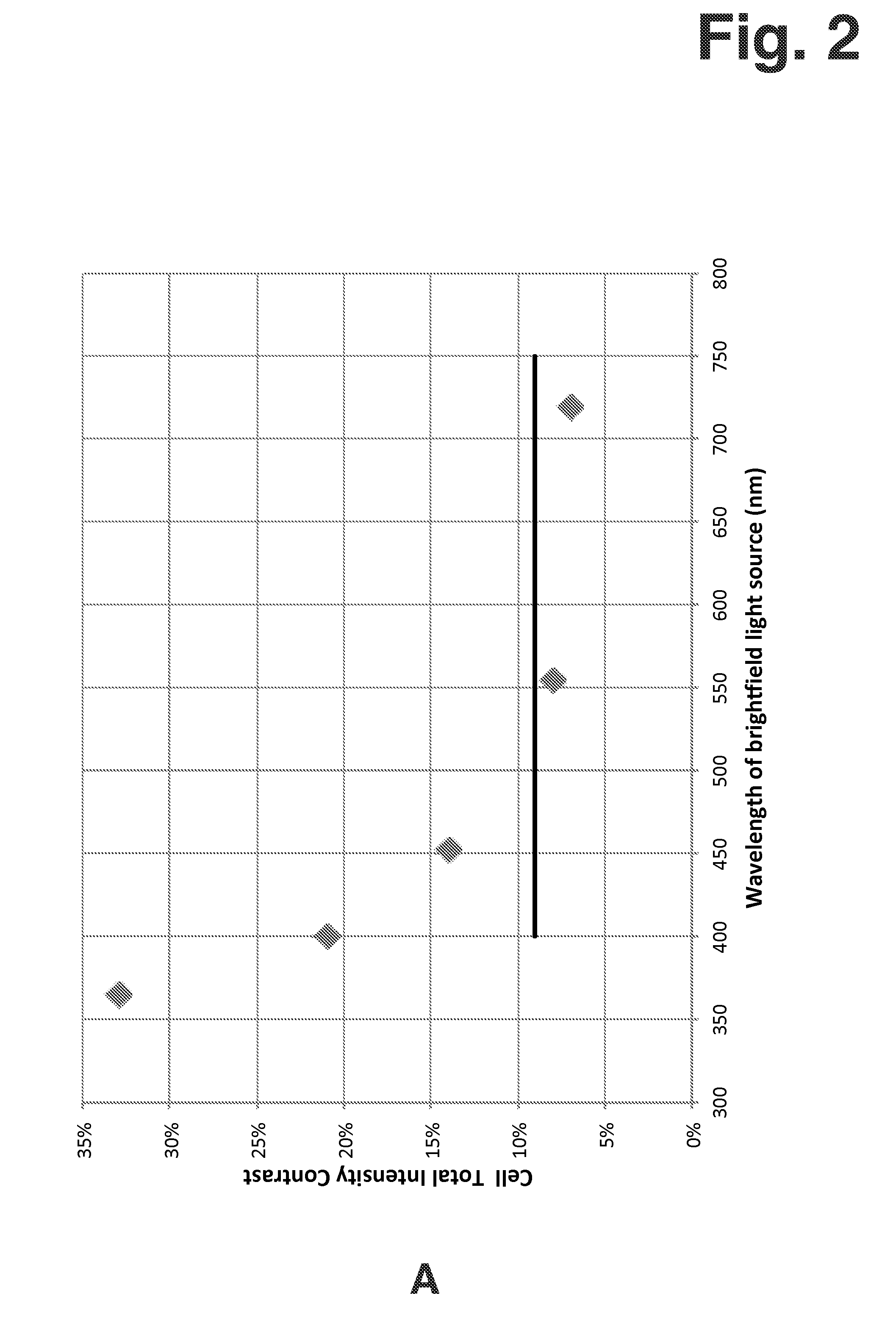
[0119]Contrast in Bright Field microscopy according to the present invention was investigated by measuring a sample of Jurkat cells (human leukemia cell line, subclone A3, ATCC CRL-2570). The measurements were performed using four light sources of different wavelength. Three of the light sources were single colour narrow-waveband Light Emitting Diodes (LEDs) and the fourth light source was a broad-waveband white LED. All light sources were in optical arrangement where emitted light was collimated when passing the sample.
[0120]The output from the narrow-waveband LEDs was used without modification but the output from the white LED was used with modification, as well as being modified using narrow-band filters. The wide-waveband light from the white LED represents typical conditions of visible microscopy. The principle wavelengths of the narrow-waveband light used in the measurement are listed in the following Table 2-1.
TABLE 2-1List of Light Sour...
example 3
Enhancement of Signal to Background
[0125]When performing fluorescence analysis of a biological sample it is important to manage excitation and emission light in order to obtain adequate contrast in the image. There are basically two approaches to improving contrast, firstly to use an excitation filter to reduce light of wavelength longer than what is used for the excitation of fluorescence and secondly to use an emission filter to reduce light of wavelength shorter than the fluorescent light from reaching the array of active detection elements.
[0126]In order to perform high-sensitivity fluorescence analysis, where signal contrast is an important aspect, it is necessary to consider all aspects of the optical system which affect the collected intensity information. This includes aspects such as excitation intensity, auto-fluorescence of any component of the system and attenuation of optical filters. In the task of improving contrast in collecting fluorescence spatial intensity informa...
PUM
| Property | Measurement | Unit |
|---|---|---|
| wavelength | aaaaa | aaaaa |
| diameter | aaaaa | aaaaa |
| width | aaaaa | aaaaa |
Abstract
Description
Claims
Application Information
 - R&D
- Intellectual Property
- Life Sciences
- Materials
- Tech Scout
- Unparalleled Data Quality
- Higher Quality Content
- 60% Fewer Hallucinations
Browse by: Latest US Patents, China's latest patents, Technical Efficacy Thesaurus, Application Domain, Technology Topic, Popular Technical Reports.
© 2025 PatSnap. All rights reserved.Legal|Privacy policy|Modern Slavery Act Transparency Statement|Sitemap|About US| Contact US: help@patsnap.com


