Compositions and Methods to Image and Quantify Inflammation

a technology of inflammation and composition, applied in the field of compositions and techniques for assessing inflammation, can solve the problems of tissue inflammation, limited noninvasive monitoring technology, and inability to accurately predict the location and quantity of leukocytes in vivo

Inactive Publication Date: 2016-10-13
CELSENSE
View PDF0 Cites 0 Cited by
  • Summary
  • Abstract
  • Description
  • Claims
  • Application Information

AI Technical Summary

Problems solved by technology

Although migration of leukocytes plays a key role in numerous diseases, present technologies available for noninvasively monitoring the location and quantity of leukocytes in vivo are limited.
The tissue sampling process is invasive and a large number of samples may be needed to ascertain the macroscopic location and extent of cellular infiltration.
Repetitive sampling of an inflamed tissue may result in further inflammation of the tissue, confounding results obtained by the process.
If untreated, the disease may cause lasting or permanent damage to the heart.
This is a dangerous procedure and subject to error due an observed inhomogeneity of leukocytes on the surface of the organ.
Existing instruments for non-invasive analysis of the trafficking of leukocytes are ill-suited for clinical use.
Light-based imaging technologies, such as bioluminescence (e.g., luciferases) technologies, are often ineffective at visualizing deep structures because most mammalian tissues are optically opaque.
Radioactive probes used in Positron Emission Tomography (PET) and / or Single Photon Emission Computed Tomography (SPECT) are highly sensitive, but the spatial resolution of cellular location is limited.
These techniques must be combined with either CT or MRI to place the detected leukocytes into anatomical context, requiring more complicated analysis and / or instrumentation.
Furthermore, longitudinal analysis of leukocyte location is limited by the time scale of the radioactive decay of the probe.

Method used


Image

  • Compositions and Methods to Image and Quantify Inflammation
  • Compositions and Methods to Image and Quantify Inflammation
  • Compositions and Methods to Image and Quantify Inflammation

Examples


Embodiment Construction

[0021]It is to be understood that the figures and descriptions of the present invention have been simplified to illustrate elements that are relevant for a clear understanding of the invention, while eliminating, for purposes of clarity, other elements that may be well known. The detailed description will be provided herein below with reference to the attached drawings.

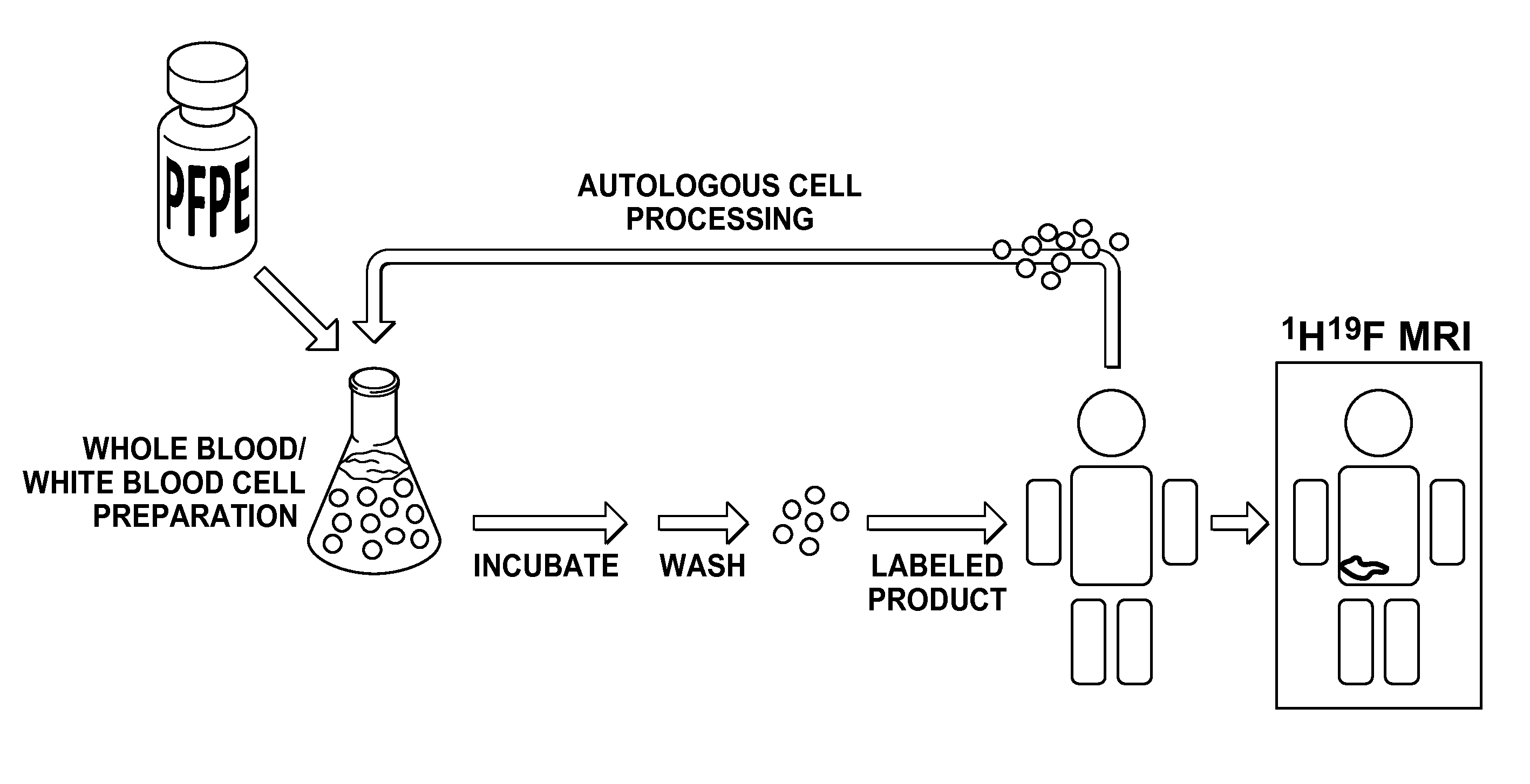
[0022]This disclosure discloses a novel method of non-invasively assessing inflammation in a patient. A schematic of the general methods of the present invention is depicted in FIG. 1. As part of the methods of the present invention, a portion of a subject's leukocytes are removed from the subject, labelled with an agent detectable in 19F MRI, and re-injected into the subject. After some time, the subject, or some portion thereof, is interrogated using 19F MRI where the labelled cells are rendered distinct from the subject. The labelled cells serve as a proxy measure of the trafficking of leukocytes in a subject. In c...


PUM

PropertyMeasurementUnit
particle sizeaaaaaaaaaa

Abstract

Compositions and methods for assessing inflammation in a subject. The present disclosure provides compositions for labeling leukocytes with a 19F-containing perfluoropolyether molecule ex vivo. In some examples, the leukocytes are obtained from the patient, enriched in a whole blood fraction, and then labeled. The labeled cells may be re-introduced into the patient. The leukocytes may accumulate at a site of inflammation, thus permitting non-invasive evaluation of inflammation in patients. The present methods provide a tool for assessing inflammation in a wide variety of autoimmune diseases and may have particular utility in intestinal diseases such as Crohn's disease, ulcerative colitis, inflammatory bowel disease, and cardio myositis.

Description

CROSS-REFERENCE TO RELATED APPLICATIONS[0001]This application claims the benefit under 35 U.S.C. § 119(e) of the earlier filing date of U.S. Provisional Patent Application No. 61 / 912,832 filed on Dec. 6, 2013 and U.S. Provisional Patent Application No. 61 / 920,498 filed on Dec. 24, 2013.BACKGROUND OF THE INVENTION[0002]1. Field of the Invention[0003]The present invention relates generally to compositions and techniques for assessing inflammation. More particularly, the present invention is directed to compositions useful in conducting magnetic resonance imaging (MRI)-based assessment of inflammation.[0004]2. Description of the Background[0005]Many biological processes are carried out by populations of cells. For example, cells of the immune system are recruited from the body to areas of injury or infection, resulting in an accumulation of leukocytes at the affected site. A marked infiltration of leukocytes often occurs in tissues affected by autoimmune diseases, physical injury, canc...

Claims


Application Information

Patent Timeline
Patent Type & Authority Applications(United States)
IPC IPC(8): A61K49/12A61K49/18
CPCA61K49/1896A61K49/126A61K49/12A61P29/00A61B5/055A61K51/0423A61K51/1203
Inventor HELFER, BROOKE M.BALDUCCI, ANTHONY G.O'HANLON, III, CHARLES F.
Owner CELSENSE
Why Eureka
Try Eureka