System and method for generating magnetic resonance imaging (MRI) images using structures of the images

a technology of magnetic resonance imaging and image structure, applied in the field of quantitative analysis of medical images, can solve the problems of limited spatial resolution, limited image resolution, and compromise of spatial resolution, and achieve the effect of less errors and less errors

Inactive Publication Date: 2017-01-05
THE GENERAL HOSPITAL CORP
View PDF1 Cites 10 Cited by
  • Summary
  • Abstract
  • Description
  • Claims
  • Application Information

AI Technical Summary

Benefits of technology

[0006]The present invention overcomes the aforementioned drawbacks by providing a system and method to interpolate low resolution (LR) images of a first contrast with the help of high resolution (HR) images of a second contrast using anatomical structures in both sets of images. To preserve the contrast in the LR images of the first contrast but prevent the dominance of the errors in them due to their low resolution, the weights used to express the interpolated images can, optionally, be calculated more than once. In the first time, the LR images may be interpolated to the high resolution and HR images of the first contrast are generated. The two sets of HR images are then coregistered. Weights are calculated using the laminar structures of the coregistered HR images of the second contrast, and then are used to generate HR images of the first contrast with less errors. In the second time, weights are calculated again using the laminar structures of both the coregistered HR images of the second contrast and the generated HR images of the first contrast with less errors, and then may be used to generated final interpolated images. In one configuration, the laminar structures are represented with feature vectors comprising image intensities, edges, and propagated edge information and weights are calculated based on the probabilities that neighbors of a given voxel have similar feature vectors to the given voxel.

Problems solved by technology

In magnetic resonance imaging (MRI), the acquisition time, short physiological phenomena, and organ motion may limit the image resolution.
In some cases, spatial resolution is limited by image acquisition time.
Thus, spatial resolution may be compromised to achieve reasonable scan time and minimize the likelihood of motion artifact.
The quality of reconstruction, however, is limited resulting in poor quality MR images.
Unfortunately, guessing or interpolation techniques often inject further errors into the process, which can degrade the quality and value of the images clinically.

Method used


Image

  • System and method for generating magnetic resonance imaging (MRI) images using structures of the images
  • System and method for generating magnetic resonance imaging (MRI) images using structures of the images
  • System and method for generating magnetic resonance imaging (MRI) images using structures of the images

Examples


Embodiment Construction

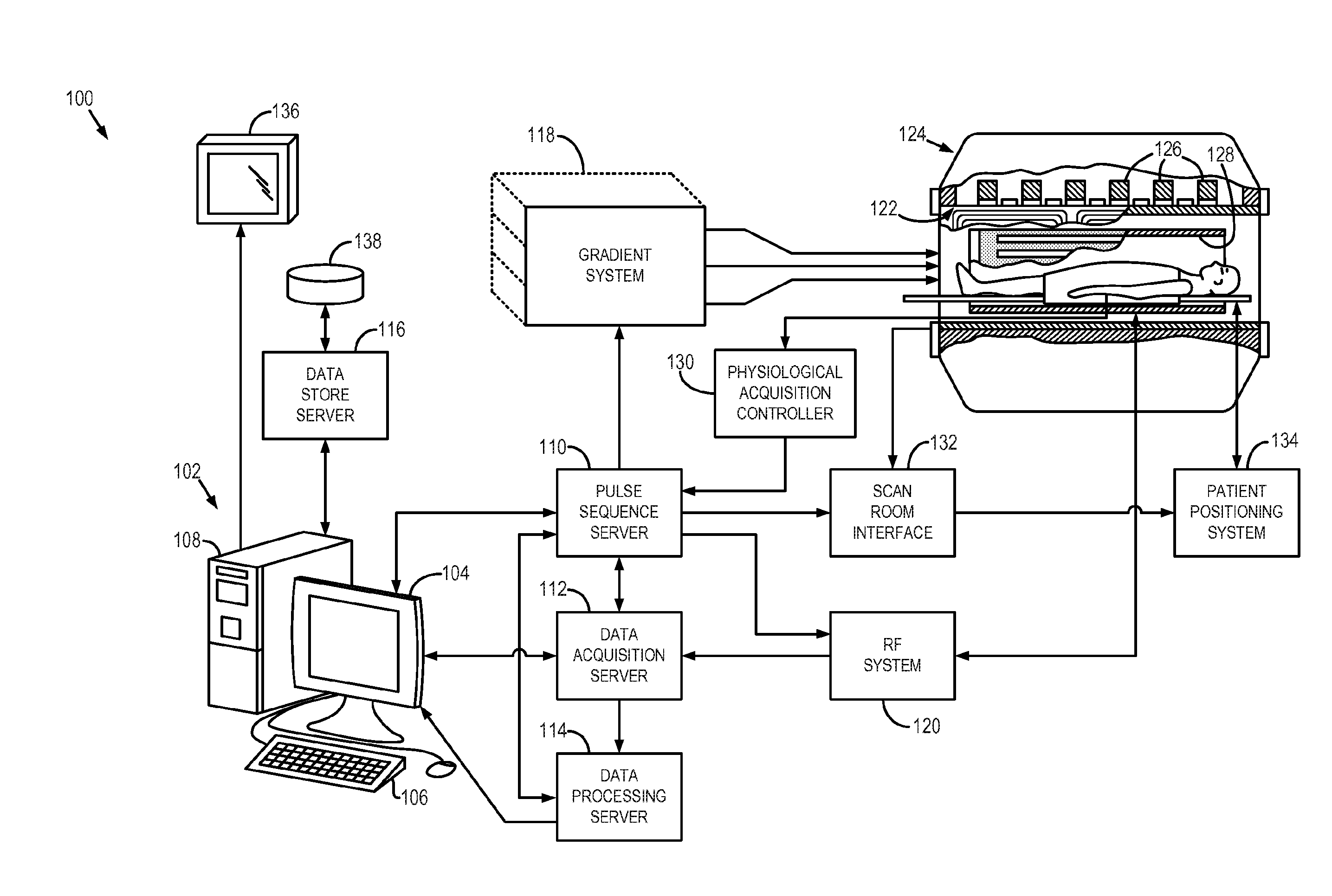
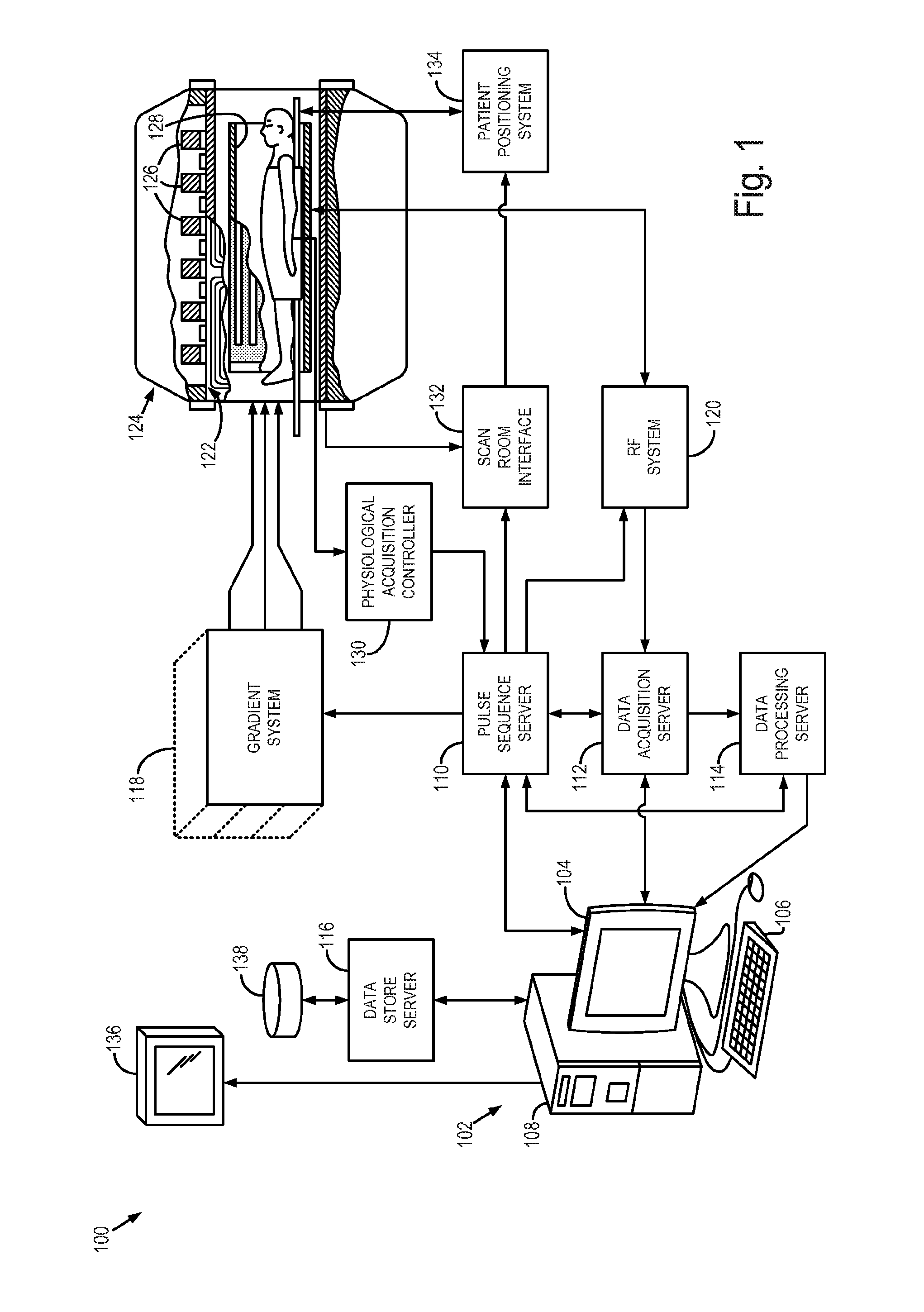
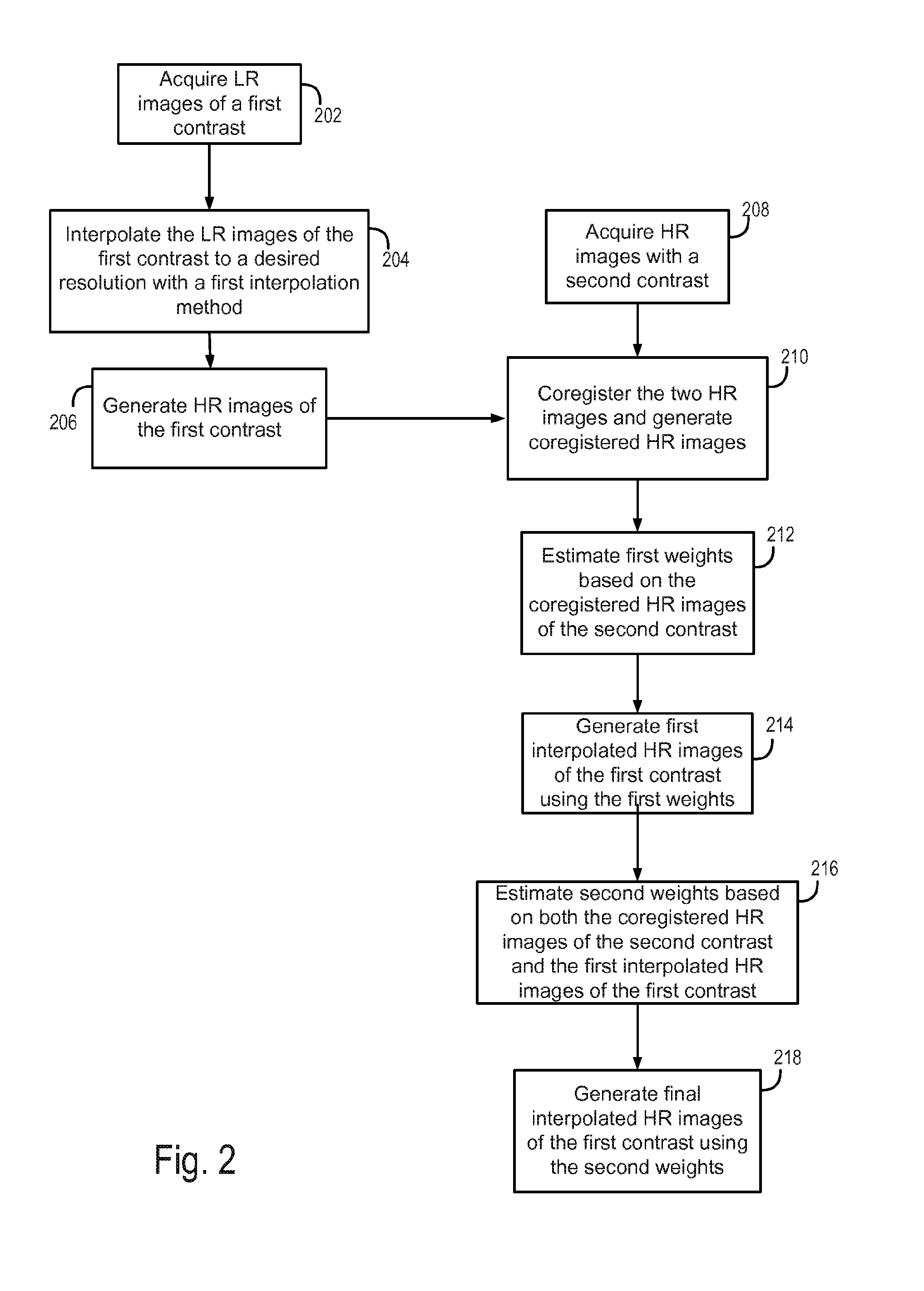
[0028]As discussed, quantitative analyses of these low resolution (LR) images are often distorted when images are transformed into a different space. For instance, voxel-wise analysis usually requires all image volumes of one subject to be aligned and analyzed in one space. Thus, if slices of different image volumes are not already acquired exactly from the same location, they need to be coregistered. Coregistration, in this case, involves guessing or interpolation. However, standard interpolation techniques are not accurate and result in distorted edges in the planes perpendicular to the acquisition plane. The reason for an inaccurate interpolation is that the neighboring voxels, from which the intensity of an unknown voxel is estimated, may not have the same tissue type as the unknown voxel. This results in an incorrect estimation of the voxel intensity using standard interpolation techniques.

[0029]As will be described, the present disclosure provides a system and method for gener...


PUM


Abstract

A system and method for generating high resolution (HR) images from low resolution (LR) images or data by selectively choosing neighbors and the tissue types of the neighbors when estimating the image intensity of a voxel with the values of the neighbors. The system and method may interpolate LR images of a first contrast with the help of the high resolution HR images of a second contrast using the anatomical structures in both sets of images.

Description

CROSS-REFERENCE TO RELATED APPLICATIONS[0001]This application claims the benefit of, and herein incorporates by reference in its entirety, U.S. Provisional Patent Application Ser. No. 61 / 930,716, filed on Jan. 23, 2014, and entitled “Upsampling MRI Images.”STATEMENT REGARDING FEDERALLY SPONSORED RESEARCH[0002]This invention was made with government support under R90-DA023427, 5U01CA154601-03, 2P41EB015896-16, R01CA129371, and K24CA125440A awarded by the National Institutes of Health. The government has certain rights in the invention.BACKGROUND OF THE INVENTION[0003]In quantitative analysis of medical images, certain spatial resolution is desired. In magnetic resonance imaging (MRI), the acquisition time, short physiological phenomena, and organ motion may limit the image resolution. For example, in dynamic contrast enhanced (DCE) and dynamic susceptibility contrast (DSC) MRI of brain, a tracer is injected into the vein during continuous rapid brain imaging. As the arrival of the bo...

Claims


Application Information

Patent Timeline
Patent Type & Authority Applications(United States)
IPC IPC(8): G01R33/56G06T7/00A61B5/00A61B5/0402A61B5/08G06T11/00G01R33/567
CPCA61B5/055G06T2207/10088G06T7/13G01R33/5608A61B5/08A61B5/7285G01R33/5601G01R33/5673G06T7/0012G06T11/003G06T2207/30016
Inventor JAFARI-LHOUZANI, KOUROSH
Owner THE GENERAL HOSPITAL CORP
Why Eureka
Try Eureka