Image-Guided Radiation Therapy

Active Publication Date: 2017-02-02
ELEKTA AB
View PDF4 Cites 16 Cited by
  • Summary
  • Abstract
  • Description
  • Claims
  • Application Information

AI Technical Summary

Benefits of technology

[0013]The present invention therefore provides, in one aspect, a radiotherapeutic apparatus for delivering a prescribed dose of radiotherapeutic radiation to a target region of patient tissue comprising a source for producing a directable beam of therapeutic radiation, wherein an image depicting patient anatomical data of at least the target region has been acquired and the prescribed dose has been mapped onto the image as the desired dose distribution to be achieved in the target region, the apparatus being adapted and configured to:
[0014]a) determine at least one beam angle corresponding to a direction from which the radiotherapeutic radiation is to be delivered from the source to the target region, and for each beam angle create a mask defi

Problems solved by technology

It is known that exposure of human or animal tissue to ionising radiation will damage the cells thus exposed.
In conventional radiation therapy, large volumes of healthy tissue can thus be exposed to harmful doses of radiation, potentially resulting in unacceptable side-effects.
However, the high energy associated with therapeutic radiation is not ideal for imaging purposes as the attenuation coefficients of the various tissue types within a patient are similar at this energy level, leading to poor image contrast.
In addition, this method is inherently two-dimensional because in conventional radiotherapy the megavoltage beams are directed at the patient from typically at least three angles, which may be insufficient to provide three-dimensional imaging.
One problem with existing and planned radiotherapy systems is to ensure that the radiation distribution is applied accurately according to the treatment plan, in terms of both the locations the radiation is delivered to and the amount of radiation delivered (the “dose”) to any particular location.
A second problem is that of beam-angle optimisation, which is to determine the “optimal” number and values of gantry angles, which is often form

Method used


Image

  • Image-Guided Radiation Therapy
  • Image-Guided Radiation Therapy
  • Image-Guided Radiation Therapy

Examples


example

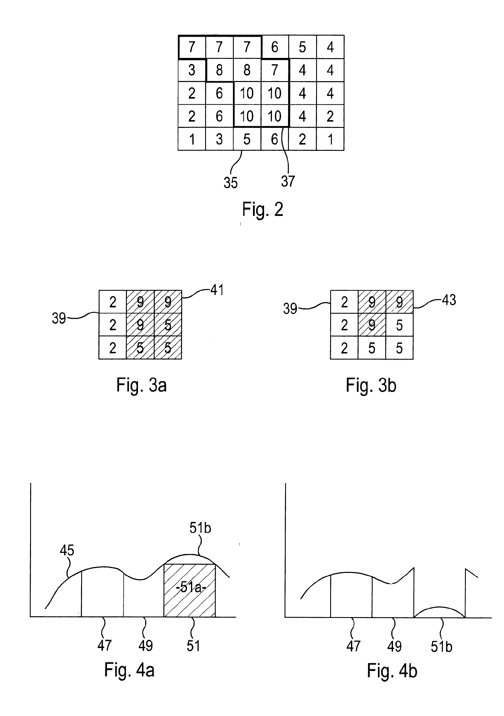
[0088]Method: Pencil beams were generated for each beam angle of the treatment and a fluence optimization performed. The pencil beams, together with the patient anatomy and the above optimal fluence form the input of the algorithm. During each of a number of iterations the following steps were performed: A fluence optimization was carried out, and each beam's fluence then split to discrete intensity levels. Deliverable segments were calculated for each of these. Each segment's area was multiplied by its intensity to determine its efficiency. The most efficient segment among all beams was then chosen to deliver a part of the calculated fluence, and the dose that would be delivered by this segment calculated. This delivered dose was then subtracted from the remaining dose. This loop was repeated until 90% of the dose had been delivered and a final segment weight optimization was performed to reach full convergence.

[0089]Results: The algorithm was tested in several prostate cases. All ...


PUM


Abstract

The disclosed systems and methods for Image-Guided Radiation Therapy (IGRT), utilises an iterative approach which adjusts a treatment plan based on inter- or intra-fraction images to improve the accuracy of the radiation delivered during the overall treatment. The prescribed dose of radiotherapeutic radiation is mapped onto the patient's anatomy using an image acquired of the region, which is to be the target for radiotherapeutic radiation. Following beam-angle-optimisation, fluence optimisation and segmentation, the efficiency of delivery of each segment is determined using an objective function, and the segments ranked according to their efficiency. The plan proceeds with the choice of the most efficient segment (or segments) to be delivered first. When this radiation has been delivered, having been tracked to establish its distribution, this delivered distribution can be subtracted from the original prescribed dose and the process repeated so that the delivered radiation gradually converges on the original prescribed dose.

Description

FIELD OF THE INVENTION[0001]The present invention relates to systems and methods for Image-Guided Radiation Therapy (IGRT), particularly but not exclusively when practised with a magnetic resonance imaging (MRI) system, such as an MRI / Linear Accelerator, or “MRL”.BACKGROUND ART[0002]IGRT is the process of frequent two and / or three-dimensional imaging, during radiation treatment, used to direct the delivery of the therapeutic radiation.[0003]It is known that exposure of human or animal tissue to ionising radiation will damage the cells thus exposed. This finds application in the treatment of pathological cells, for example. In order to treat tumours deep within the body of the patient, the radiation must however penetrate the healthy tissue in order to irradiate and destroy the pathological cells. In conventional radiation therapy, large volumes of healthy tissue can thus be exposed to harmful doses of radiation, potentially resulting in unacceptable side-effects. It is therefore des...

Claims


Application Information

Patent Timeline
IPC IPC(8): A61N5/10
CPCA61N5/1067A61N5/1047A61N5/1031A61N5/1039A61N2005/1055A61N5/1038A61N5/1049A61N2005/1034A61N2005/1054A61N5/1071A61N5/1037A61N2005/1058
Inventor KONTAXIS, CHARISBOL, GIJSBERT HERMANRAAYMAKERS, BAS WILLEM
Owner ELEKTA AB
Why Eureka
Try Eureka