
Intra-operative medical image viewing system and method

a medical image and viewing system technology, applied in the field of intraoperative medical image viewing system and method, can solve the problems of increasing affecting the patient's recovery, and difficult to eradicate, so as to achieve easy and intuitive implementation, and reduce the risk of a tiny hand movemen

Inactive Publication Date: 2017-02-16
SURGERATI
View PDF3 Cites 58 Cited by
  • Summary
  • Abstract
  • Description
  • Claims
  • Application Information

AI Technical Summary

Benefits of technology

The invention makes it easier for surgeons to view medical images during operations without having to look away from the patient or sustain heavy cognitive loading. This reduces distraction and fatigue, leading to greater efficiency and productivity. The invention also helps surgeons learn faster and perform better, resulting in better effectiveness.

Problems solved by technology

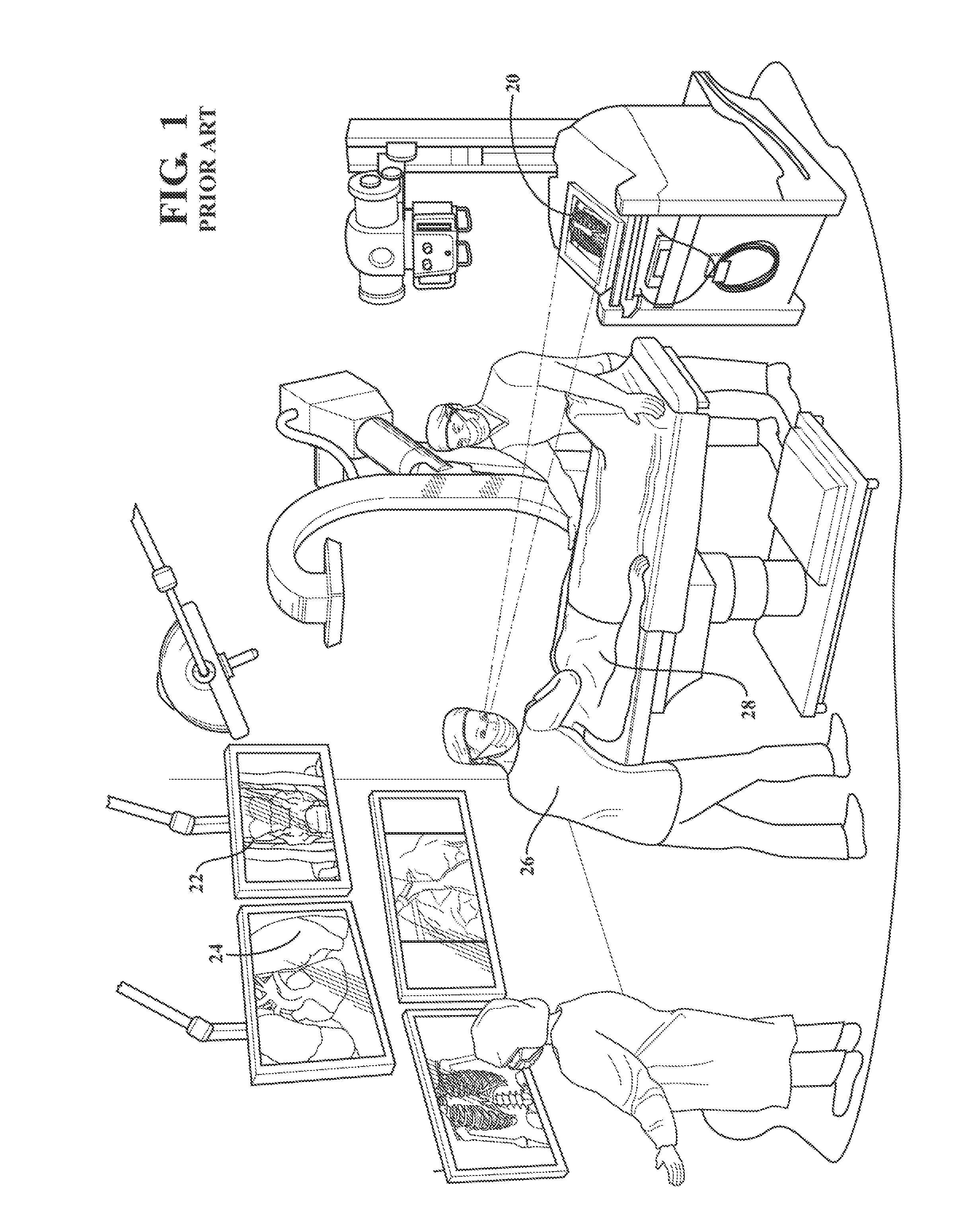
An operating room that is filled with many display screens all presenting different relevant anatomical or pathological image data to the surgeon causes several problems in the medical community, which problems have proven particularly difficult to eradicate.
A first problem relates to distraction of the surgeon's attention posed by the need to frequently look away from her patient in order to see the images on one or more display screens dispersed about the operating room.
The risk of a tiny, unwanted hand movement rises each time a surgeon must consult an image on a screen that is located some distance away from the patient.
A second problem that is provoked by the presence of multiple display screens in an operating room relates to compounding a surgeon's cognitive load.
While image registering a single static image to the patient may not be particularly taxing, the cognitive load quickly compounds when there are many display screens to be consulted, each exhibiting an image taken from yet a different perspective or presented in a different scale.
Yet another problem that is provoked by the presence of multiple display screens in an operating room relates to ergonomics.
Namely, the occupational safety and health of a surgeon is directly compromised by the required use of many widely-dispersed images during a surgical procedure.
Surgery does not afford the practitioner with the ability to rest or change positions at will in order to combat muscle cramps or nerve aggravations.
On a daily basis, this physical fatigue limits a surgeon's ability to perform at optimum ability during long shifts.
Over time, the stresses placed on the surgeon accumulate to the point where the injuries accumulate / compound and become chronic and must either be remediated through medical intervention or the surgeon prematurely limits (or truncates) her service career.
Furthermore, these problems can be inter-related.
Issues associated with cognitive load and ergonomics compound each other to diminish a surgeon's working efficiency, which affect the patient by increasing the length of time they must undergo a surgical procedure.
Naturally, increased procedure time impacts the surgeon's health but also the surgeon's productivity.
That is, with more time in each surgery the surgeon can do fewer operations over the course of a year, which also then limits the surgeon's ability to gain experience.
Increased procedure time impacts the patient in a number of ways also, including increased risks associated with prolonged time under anesthesia and its after-affects, increased risk for infections attributed to longer open incision times, longer hospital stays, increased medical costs, and the like.
Finding a solution to these persistent image-related problems in the operating room has been elusive.
That is to say, a solution that works only in the lab or only for a small sub-set of practitioners will not be genuinely viable as a marketable product.
But not all surgeons are alike, and what may be regarded by one surgeon as practical will be deemed unacceptably impractical by another.
It is predictable that a majority of surgeons will not adopt a solution if the solution is perceived to be overly complicated or as requiring a high degree of training to master.
A degenerative spiral results, because the current state of the art has no simpler, more intuitive option than adding more display screens to exhibit patient medical images in an operating room.

Method used


Image

  • Intra-operative medical image viewing system and method
  • Intra-operative medical image viewing system and method
  • Intra-operative medical image viewing system and method

Examples


Embodiment Construction

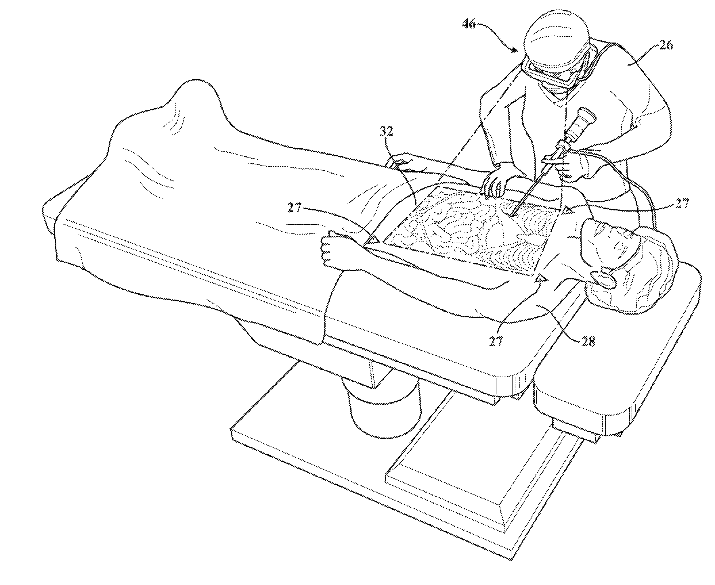
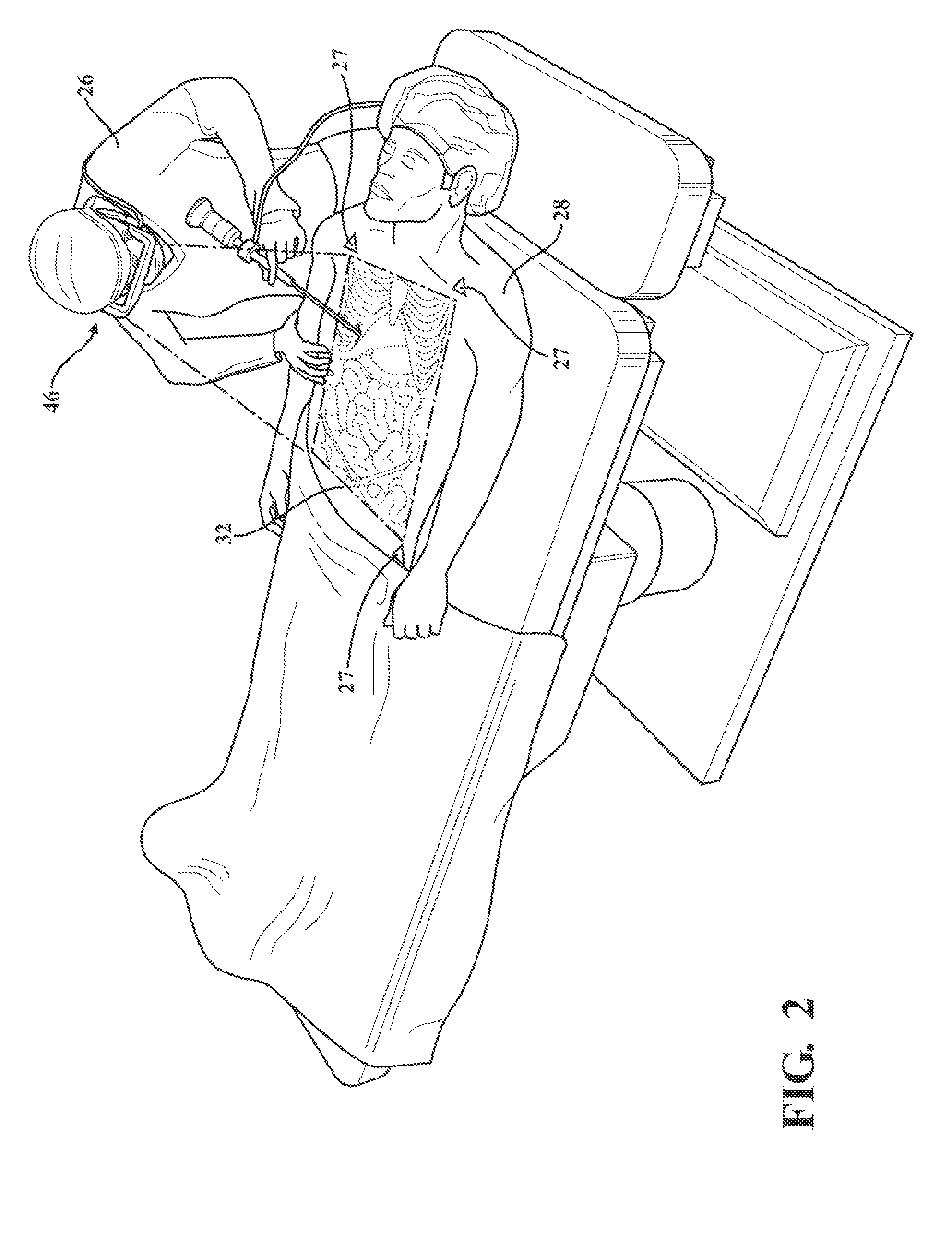
[0032]The exemplary embodiment can provide an intra-operative medical image viewing system 34 and method for displaying and interacting with two-dimensional, 2-½-Dimentional, or three-dimensional visual data in real-time and in perceived three-dimensional space. The system 34 can present a selectively or variably transparent image of an anatomical feature of a patient 28 to a surgeon 26 during surgery, as the surgeon 26 maintains a viewing perspective generally centered on the actual anatomical feature of the patient 28 or at least toward the patient 28 on whom some operation is being performed. The image as perceived by the surgeon 26 is selectively and / or variably transparent, in the sense that the surgeon 26 controls the image opacity throughout the range of fully transparent, e.g., when the image is not in use, to fully opaque, e.g., when high-contrast is desired, and through some if not all levels in-between. In most cases, the medical image appears to the surgeon to be located...


PUM


Abstract

An intra-operative medical image viewing system can allow a surgeon to maintain a viewing perspective on the patient while calling-up visual images on-the-fly. A digital image source has at least one image file representative of an anatomical or pathological feature of a patient. A display is worn by the surgeon or positioned between the surgeon and her patient during surgery. The display is selectively transparent, and exhibits to the surgeon an image derived from the image file. An image control unit retrieves the image file from the image source and controls the display so that at least a portion of the image depiction can be exhibited and modified at will by the surgeon. A plurality of peripheral devices are each configured to receive an image control input from the surgeon and, in response, generate an image control signal. Each peripheral accepts a different user-interface modality.

Description

CROSS REFERENCE TO RELATED APPLICATIONS[0001]This application claims priority to Provisional Patent Application No. 61 / 982,787 filed Apr. 22, 2014, the entire disclosure of which is hereby incorporated by reference and relied upon.BACKGROUND OF THE INVENTION[0002]Field of the Invention[0003]The invention relates generally to generating, processing, transmitting or transiently displaying images in a medical environment, in which the local light variations composing the images may change with time, and more particularly to subject matter in which the image includes portions indicating the three-dimensional nature of the original object.[0004]Description of Related Art[0005]In a surgical environment, there are often many display screens each displaying different visual information that is of interest to the medical practitioner, such as a surgeon. In particular, the visual information may include images representing an anatomical or pathological feature of a patient, such as an X-ray, ...

Claims


Application Information

Patent Timeline
IPC IPC(8): A61B90/00H04N13/04
CPCA61B90/37H04N13/044H04N13/0497A61B2017/00203A61B2017/00207A61B2017/00212A61B2090/502A61B2034/2065A61B2090/366A61B2090/367A61B2090/371A61B2090/372A61B2034/2048H04N13/344H04N13/398
Inventor DOO, FLORENCE XINIBLOOM, DAVID C.
Owner SURGERATI
Learn More