
Methods and probes for vaginal tactile and electromyographic imaging and location-guided female pelvic floor therapy

a technology of electromyographic imaging and probes, applied in the field of probes for vaginal tactile and electromyographic imaging and location-guided female pelvic floor therapy, can solve the problems of significant morbidity and quality of life impact, physical training and medication are often not effective in treating ui, and lack of objective functional and anatomically specific guidance to select precise locations for effective therapy

Active Publication Date: 2017-03-09
ADVANCED TACTILE IMAGING
View PDF2 Cites 7 Cited by
  • Summary
  • Abstract
  • Description
  • Claims
  • Application Information

AI Technical Summary

Benefits of technology

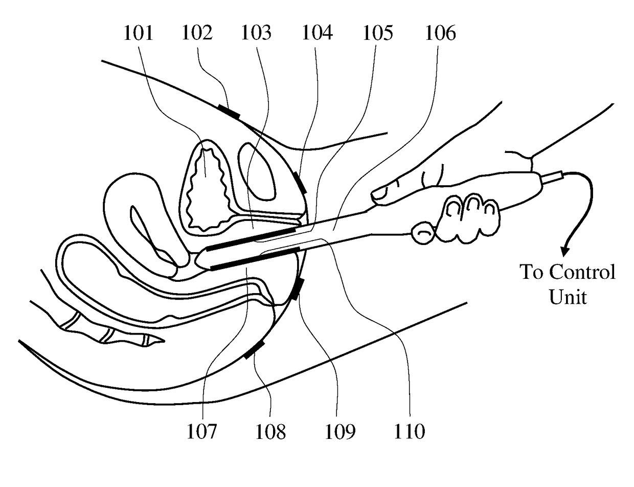
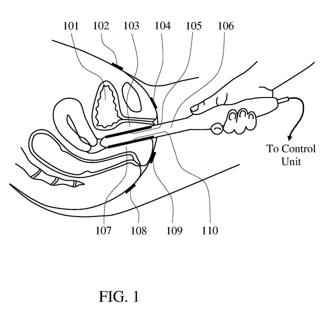
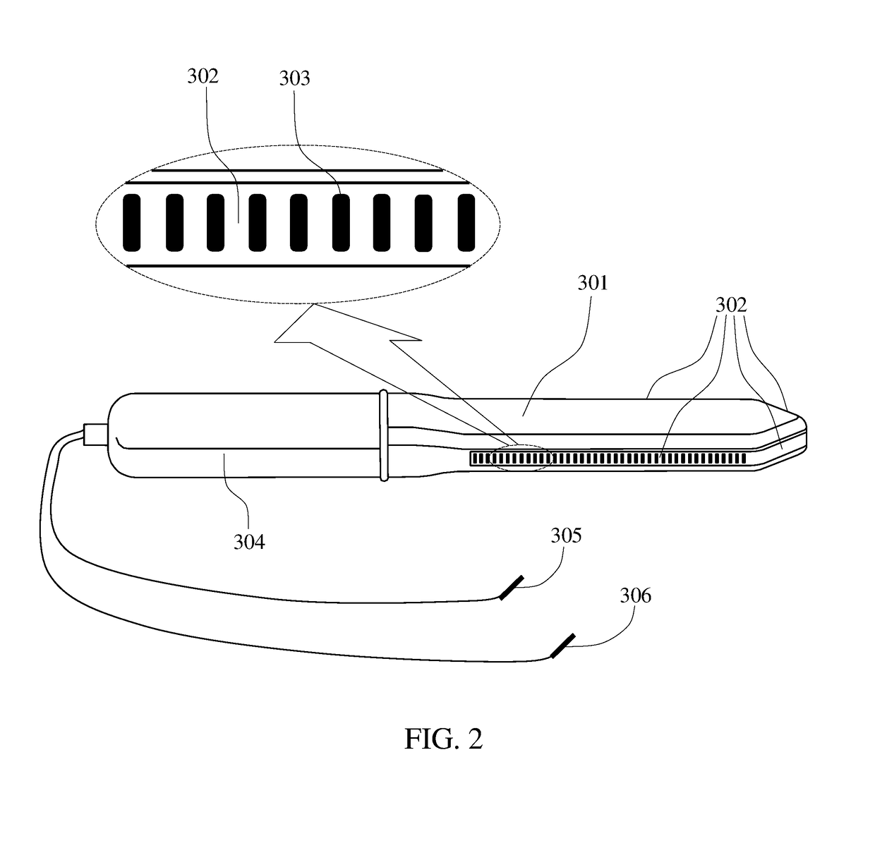
The present invention provides methods and devices for objective characterization of the female pelvic floor conditions. The invention aims to overcome the drawbacks of prior art methods and provide real-time visualization of pelvic floor muscles' contractile capabilities and electromyographic activities. The invention also allows for the detection and treatment of damaged or weak pelvic floor muscles by measuring a tactile response and an electromyographic response during pelvic floor muscles contractions. The invention also provides methods and devices for location-guided therapy of damaged or weak pelvic floor muscles using electrical stimulation or drug injection. The vaginal probe used in the invention has a front portion for atraumatic insertion into the vaginal canal and includes a plurality of tactile sensors and electrodes for recording the tactile and electromyographic responses of pelvic floor muscles during contraction. The invention also provides reference electrodes for attachment to the skin near the vaginal canal.

Problems solved by technology

These conditions are socially embarrassing, causing significant morbidity and quality of life impact.
However, numerous patient reports of adverse events are associated with surgical mesh; these include vaginal extrusion, erosion, infection, pain, dyspareunia, lower urinary tract symptoms, recurrence of incontinence, urinary retention, and blood vessel injury.
Physical training and medications are often not effective for treatments of UI.
The transvaginal electrical simulation demonstrated feasibility for UI and OAB treatments but lacks objective functional and anatomically-specific guidance to select precise location for the effective therapy.
To date, there is no standardized, reproducible tool to accurately assess the pelvic floor muscles function in anatomically-specific manner, and to apply guided muscle stimulation for UI and OAB therapy.
However, its effectiveness remains poorly understood with some studies suggesting that biofeedback offers no advantage over the isolated pelvic floor muscle training.
Commercially available vaginal probes designed to record EMG from the pelvic floor muscles have numerous deficiencies in their design, such as problems with probe geometry, electrode size, location, and / or configuration.
Many probe designs are prone to recording motion artifacts, crosstalk, and / or inappropriate EMG signals.
The current art in the field indicates that their measurement accuracy and reproducibility are too low for the reliable characterization of the pelvic floor muscles conditions.
Nevertheless, some controversy exists on effectiveness of EMS as a stand-alone therapy for female SUI, which may be attributed by the inability to apply guided and targeted EMS.

Method used


Image

  • Methods and probes for vaginal tactile and electromyographic imaging and location-guided female pelvic floor therapy
  • Methods and probes for vaginal tactile and electromyographic imaging and location-guided female pelvic floor therapy
  • Methods and probes for vaginal tactile and electromyographic imaging and location-guided female pelvic floor therapy

Examples


Embodiment Construction

[0035]The following description sets forth various examples along with specific details to provide a thorough understanding of claimed subject matter. It will be understood by those skilled in the art, however, that claimed subject matter may be practiced without one or more of the specific details disclosed herein. Further, in some circumstances, well-known methods, procedures, systems, components and / or circuits have not been described in detail in order to avoid unnecessarily obscuring claimed subject matter. In the following detailed description, reference is made to the accompanying drawings, which form a part hereof. In the drawings, similar symbols typically identify similar components, unless context dictates otherwise. The illustrative embodiments described in the detailed description, drawings, and claims are not meant to be limiting. Other embodiments may be utilized, and other changes may be made, without departing from the spirit or scope of the subject matter presented...


PUM


Abstract

Methods and a probes are disclosed for vaginal tactile and electromyographic imaging, and location-guided female pelvic floor therapy. Methods include the steps of inserting a vaginal probe equipped with tactile sensors and electrodes acting as electromyographic sensors into a vagina along a vaginal canal to separate apart two opposing vaginal walls, recording a tactile response and an electromyographic response from at least one of two opposing vaginal walls during pelvic floor muscle contraction, determining locations along the vaginal canal for delivery of therapy based on presence of tactile response above a predetermined tactile threshold and / or presence of electromyographic response above a predetermined EMG threshold along the vaginal probe, selecting at least one of target locations to be used for location-guided therapy, and applying the therapy such as electrical muscle stimulation to at least one of selected locations using the same electrodes at these locations. The probe comprises a probe housing, a tactile sensors array, a plurality of electrodes which can be used as an electromyography array or as an electric muscle stimulation array, and at least one reference electrode.

Description

CROSS-REFERENCE TO RELATED APPLICATIONS[0001]This patent application claims a priority benefit from the U.S. Provisional Patent Application No. 62 / 214,042 filed Sep. 3, 2015 by the same inventor and with the same title. This provisional patent application is incorporated herewith by reference in its entirety.GOVERNMENT-SUPPORTED RESEARCH[0002]This invention was made with the US Government support under grant No. R43AG053935 awarded by the National Institute on Aging. The Government has certain rights in this invention.FIELD OF THE INVENTION[0003]The present invention generally relates to methods and devices for female pelvic floor imaging and treatment. Specifically, the invention describes methods and devices for providing vaginal tactile and electromyographic imaging and location-guided female pelvic floor therapy.BACKGROUND[0004]Urinary incontinence (UI) is defined as the complaint of any involuntary loss of urine. The most common type of UI is stress urinary incontinence (SUI), ...

Claims


Application Information

Patent Timeline
Patent Type & Authority Applications(United States)
IPC IPC(8): A61B5/00A61B5/22A61B5/0488A61N1/05A61N1/36
CPCA61B5/4836A61N1/0524A61N1/36007A61B5/6847A61B5/227A61B5/4337A61B5/04882A61B5/0035A61B5/065A61B5/6885A61B5/391
Inventor EGOROV, VLADIMIR
Owner ADVANCED TACTILE IMAGING
Learn More