Hybridoma cell lines (my-c-cc0c2-235-3h8) and use thereof for producing a monoclonal antibody against the human cardiac myosin binding protein c (c-protein, mybpc3, cmybp-c or my-c)

a hybridoma and antibody technology, applied in the field of mouse hybridoma clone producing monoclonal antibodies, can solve the problems of inability to detect low ctn concentrations in the first hours after symptoms begin to manifest, lack of analytical sensitivity for detecting low ctn concentrations in the first hours, etc., and achieve the effect of early diagnosis of cardiac infarction

Active Publication Date: 2017-03-30
KINGS COLLEGE LONDON
View PDF0 Cites 1 Cited by
  • Summary
  • Abstract
  • Description
  • Claims
  • Application Information

AI Technical Summary

Benefits of technology

[0013]The object is achieved by generating a hybridoma cell clone producing a monoclonal antibody that detects and binds a cardiac epitope in the My-C, and that has no cross-reactivity with respect to the myosin-binding proteins of the skeletal muscles. The hybridoma cell line can be obtained by fusing myeloma cells with spleen cells of a test animal, in particular a mouse, immunized against recombinant My-C. The hybridoma cell line was deposited on Dec. 10, 2013 with the DSMZ with accession number DSM ACC3223 in accordance with the requirements of the Budapest Treaty. The antibody produced by this hybridoma cell clone, in combination with one or more other monoclonal antibodies in an ELISA, is suitable for sensitively determining the concentration of My-C in the serum, and thus for the early diagnosis of cardiac infarctions.

Problems solved by technology

[2] Cardiac troponins (cTn), however, have disadvantages, and new biomarkers could prove to be very valuable.
The cTn concentration in the serum reaches a maximum only 16 to 18 hours after the onset of symptoms, and one disadvantage of the existing cTn tests is the lack of analytical sensitivity for detecting low cTn concentrations in the first hours after symptoms begin to manifest.
While a number of biomarkers exist that are released more quickly after an infarction, none of these have prevailed, since these are not expressed in a cardioselective manner.

Method used


Image

  • Hybridoma cell lines (my-c-cc0c2-235-3h8) and use thereof for producing a monoclonal antibody against the human cardiac myosin binding protein c (c-protein, mybpc3, cmybp-c or my-c)
  • Hybridoma cell lines (my-c-cc0c2-235-3h8) and use thereof for producing a monoclonal antibody against the human cardiac myosin binding protein c (c-protein, mybpc3, cmybp-c or my-c)
  • Hybridoma cell lines (my-c-cc0c2-235-3h8) and use thereof for producing a monoclonal antibody against the human cardiac myosin binding protein c (c-protein, mybpc3, cmybp-c or my-c)

Examples


example 1

Production of the Hybridoma Cell Line

[0028]The spleen of a mouse immunized in the known manner with cC0C2 of the My-C is removed under sterile conditions, and the spleen cells are flushed out of the spleen capsule using RPMI 1640 medium (Life Technologies™, Karlsruhe) with a syringe and isolated. The spleen cells are pelletized (10 minutes at 300×g), washed three times with RPMI 1640 medium, and resuspended in RPMI 1640 medium. They are then fused with myeloma cells of the line P3X63Ag8.653 (ATTC CPL 1580). For this purpose, cultivated myeloma cells, which are in the log phase of growth, are likewise pelletized and washed three times. 1×10⁸ spleen cells and 5×10⁷ myeloma cells are pipetted into a centrifuge tube, mixed intensively and centrifuged; 1.5 ml preheated 50% polyethylene glycol 1500 (Roche, Basel) is added dropwise to the cell sediment within one minute, while the tubule is continuously rotated at 37° C. The fusion batch is then incubated for another minute at 37° C. In th...

example 2

Selection of the Antibody-Producing Clones

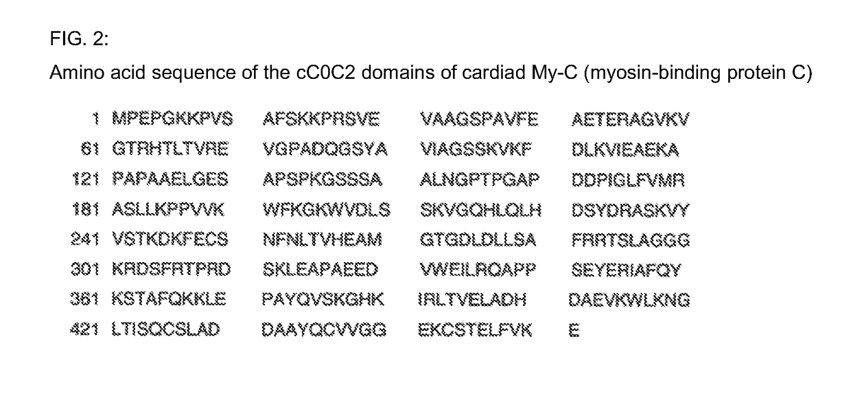
[0029]All growing clones or the antibodies thereof were tested for reactivity using an enzyme-linked immunosorbent assay (ELISA). The immunosorbent was the immunogen, this being the recombinant cC0C2 domain of the My-C (approximately 2 μg / ml).

[0030]Carrying out the ELISA:[0031]1. Coat each of the microtiter plates (Costar, high binding) with 50 μl immunogen solution per well at 4° C. over night;[0032]2. wash the microtiter plates (MTP) 3 times with Tris-buffered saline (TBS), pH 7.4;[0033]3. block the MTP using 200 μl blocking reagent (Boehringer, Mannheim) per well, 1 hour at 37° C.;[0034]4. wash the MTP 3 times with NaCl Tween 20;[0035]5. incubate with culture supernatant of the hybridoma cultures; 50 μl per well, respectively, diluted approximately 1:2 with TBS Tween 20;[0036]6. wash the MTP 3 times with NaCl Tween 20;[0037]7. incubate with anti-mouse Ig antibodies, coupled to peroxidase, 50 μl per well, 1 hour at room temperature;[0038]8...

example 3

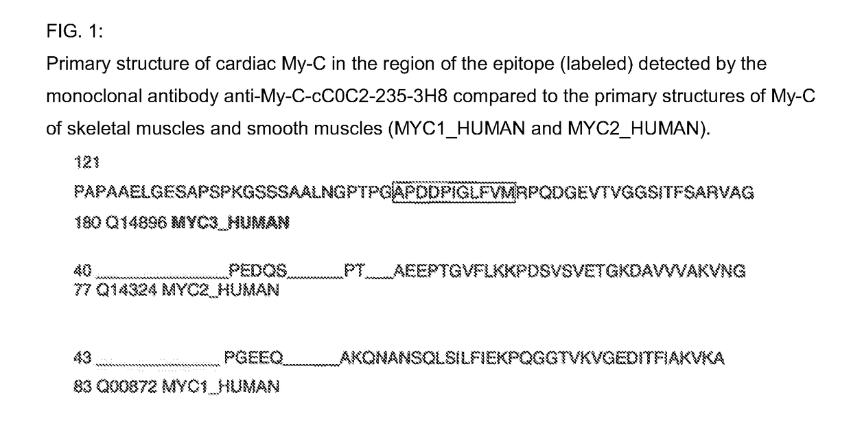
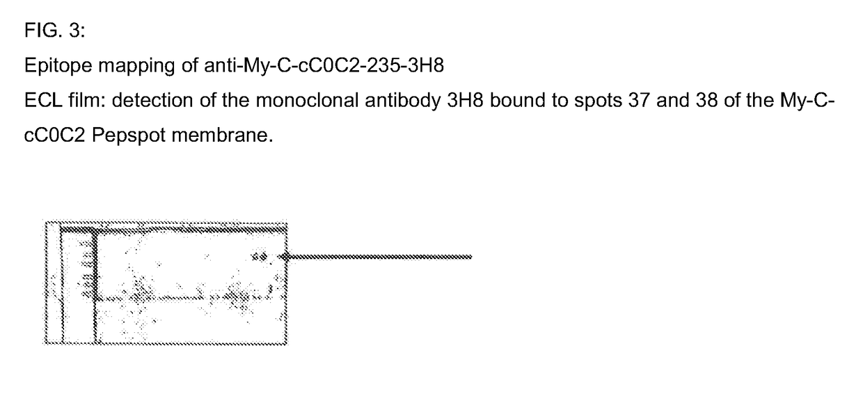
[0041]Epitope Mapping for the Monoclonal Antibody 3H8 in the Human Cardiac My-C

[0042]The binding site of the monoclonal antibody 3H8 was identified by way of the peptide scanning method. For this purpose, the entire amino acid sequence of the human cC0C2 domain of the My-C that was used for the immunization is divided into a total of 111 overlapping amino acid sequences, each having a length of 15 amino acids. These sequences are synthesized as individual peptides in spots directly on a cellulose membrane. The membrane is incubated with the antibody-containing culture supernatants of the hybridomas, and the binding sites of the antibodies are rendered visible by way of incubation with a peroxidase-coupled anti-mouse Ig antibody. For this purpose, after washing three times with TBS Tween, the membrane is placed between copy film, and then incubated for 3 minutes with the ECL™ (enhanced chemiluminescent) detection reagent (Amersham, Braunschweig). An applied film (Hyperfilm ECL™ [RPN ...


PUM

PropertyMeasurementUnit
concentrationaaaaaaaaaa
concentrationsaaaaaaaaaa
lengthaaaaaaaaaa

Abstract

Monoclonal antibodies, which can be produced in vitro, against cardiac epitopes of the human My-C are produced by generating myeloma cell clones that produce such specific antibodies having epitope specificity. These monoclonal antibodies allow, among other things, the creation of an enzyme-linked immunosorbent assay (ELISA) for the specific, cross-reactivity-free quantitative determination of My-C in serum, plasma, whole blood or other body fluid. Specifically, a hybridoma cell clone producing a monoclonal antibody that detects and binds a cardiac epitope in the My-C is produced, which has no cross-reactivity with respect to the myosin-binding proteins of the skeletal muscles. The hybridoma cell line can be obtained by fusing myeloma cells with spleen cells of a test animal, in particular a mouse, immunized against recombinant My-C. The invention furthermore relates to epitope-specific antibodies produced by the hybridoma cell line, and to the use thereof.

Description

BACKGROUND OF THE INVENTION[0001]The invention relates to a mouse hybridoma clone producing a monoclonal antibody (anti-My-C-cC0C2-235-3H8; IgG1, kappa), which is directed against the cardiac myosin-binding protein C (C-protein, MYBPC3, cMyBP-C or My-C) and detects the same, and which does not react with the closely related isomers of My-C from the skeletal muscles. This monoclonal antibody is suitable as a catcher antibody or as a detection antibody for creating an enzyme-linked immunosorbent assay (ELISA) for quantitatively determining My-C in serum, plasma, whole blood or other body fluids for the early diagnosis of cardiac infarctions. Within the scope of this diagnostic procedure, this can allow a considerably early treatment of myocardial infarctions.[0002]Due to the acute life-threatening hazard, myocardial infarctions must be diagnosed quickly and distinguished from other causes of chest pain. [1][0003]Determining biomarkers of myocardial necroses has meanwhile become an ess...

Claims


Application Information

Patent Timeline
Patent Type & Authority Applications(United States)
IPC IPC(8): C07K16/32G01N33/574
CPCC07K16/32G01N33/5748C07K2317/34C07K2317/20C07K2317/14C07K16/18G01N33/6893G01N2800/324G01N2333/4712
Inventor WEBER, EKKEHARDMEDEK, RITA
Owner KINGS COLLEGE LONDON
Why Eureka
Try Eureka