
Magnetic resonance imaging method and apparatus with simultaneous image acquisition of multiple sub-volumes with synchronous acquisition of navigators

a magnetic resonance imaging and navigator technology, applied in the field of magnetic resonance imaging apparatus, can solve the problems of loss of consistency of scanned data with the described imaging methods, long image recording time, and reduced comfort of the patient being examined, so as to improve the effect of movement correction

Active Publication Date: 2017-05-25
SIEMENS HEALTHCARE GMBH
View PDF1 Cites 17 Cited by
  • Summary
  • Abstract
  • Description
  • Claims
  • Application Information

AI Technical Summary

Benefits of technology

The present invention provides a method for controlling a magnetic resonance imaging apparatus that improves movement correction. The method involves simultaneously recording an image and a navigator volume, which can be used for movement correction of the image data. The simultaneous recording of the navigator data allows for more precise movement correction than conventional methods. The navigator data can be acquired with different contrast properties, which can affect the level of detail and impact the exposure of patients and the apparatus. The method can be easily upgraded by software and can enhance the quality of image recordings of dynamic processes. The acquired navigator image data can be used for real-time position correction and can be further used for retroactive correction to improve image quality. The navigator volume can be scanned at a higher speed or more frequently than the image volume, so it can detect and compensate for movement quickly.

Problems solved by technology

However, the image recording times are very long due to the long relaxation times of the spins, and this entails a reduction in comfort of the patient being examined.
In the case of a movement of the patient, the consistency of the scanned data is lost with the described imaging methods, and this is reflected by image artifacts in the reconstructed image.
This can be disadvantageous, for example, if the image contrast or the noise behavior is not suitable for movement detection.
In addition, it is possible that the mapped anatomy is not suitable for the chosen method of movement correction.
In general, however, this is not possible for all sequences without changing the time response, in particular it is not possible for those sequences that do not have the unused periods necessary for this.
An additional drawback of the described methods (in particular of the second group) is that the recording of the navigator image and the acquisition of the MR signal for imaging take place with at slightly staggered intervals.
Even for the case where the movement of the navigator can be exactly detected and be compensated by the sequence, resulting artifacts remain in the MR data because the movement content between the time of acquisition of the navigator and the time of acquisition of the desired MR imaging signal is not detected and therefore cannot be compensated either.

Method used


Image

  • Magnetic resonance imaging method and apparatus with simultaneous image acquisition of multiple sub-volumes with synchronous acquisition of navigators
  • Magnetic resonance imaging method and apparatus with simultaneous image acquisition of multiple sub-volumes with synchronous acquisition of navigators
  • Magnetic resonance imaging method and apparatus with simultaneous image acquisition of multiple sub-volumes with synchronous acquisition of navigators

Examples


Embodiment Construction

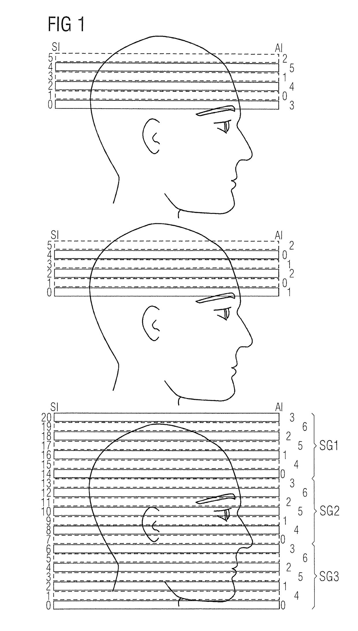
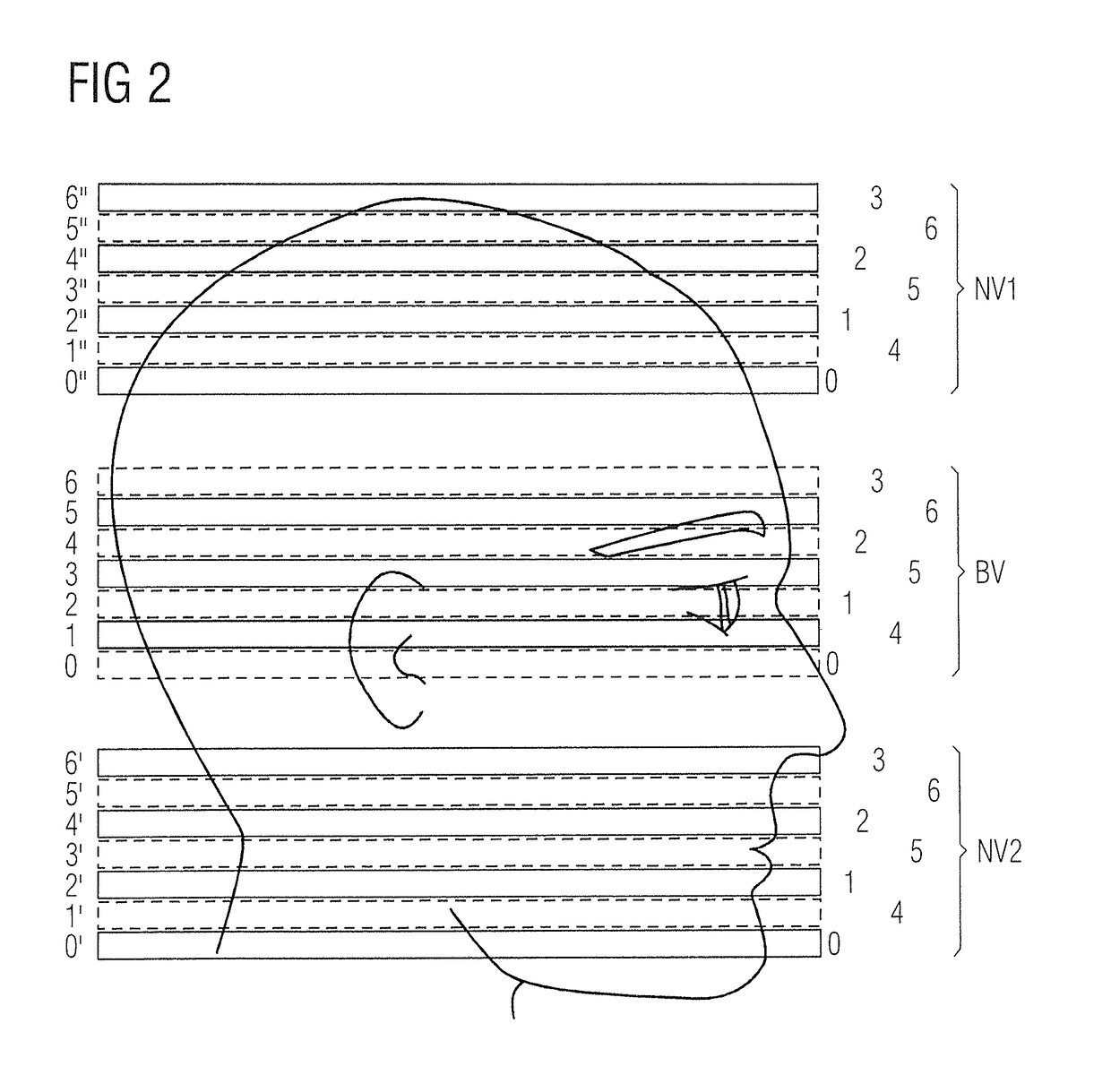
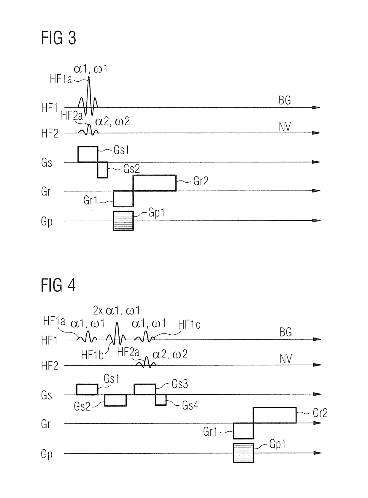
[0081]FIG. 1 shows three diagrams for illustrating an acquisition scheme for simultaneous multi-slice imaging (SMS). The top illustration shows an acquisition scheme for an MR magnetic resonance imaging method with individual slice recording. An unaccelerated scan having six slices recorded in a convoluted manner is shown. For the case of echo-planar imaging, six slice excitations and six readout cycles are required for this purpose. The left edge of the top diagram shows a slice index SI of a respective slice. The slice index SI runs in this case from 0 to 5. The right edge of the top diagram shows acquisition indices AI. These indicate the sequence in which a particular slice is excited and read out. Excitation of individual slices occurs in the top diagram takes place in a sequence that does not proceed precisely in numerical order. In other words, the slices are excited and read out in the sequence 1, 3, 5, 0, 2, 4.

[0082]In a middle image representation in FIG. 1, six slices are...


PUM


Abstract

In a magnetic resonance imaging apparatus and a method for generating magnetic resonance image data of a field of view of an examination object, magnetic resonance raw data are acquired by preferably different transverse magnetizations being excited in at least one sub-volume of a navigator volume and at least one sub-volume of an image volume, and are used for position determination and for imaging. These preferably different transverse magnetizations are simultaneously present in at least one period of the scan.

Description

BACKGROUND OF THE INVENTION[0001]Field of the Invention[0002]The invention concerns a method for controlling a magnetic resonance imaging apparatus as well as a control sequence-determining computer and a magnetic resonance imaging apparatus that implement such a method.[0003]Description of the Prior Art[0004]In medicine two-dimensional or three-dimensional image data as well as a time series of image data are often generated, using modern imaging methods, and such data are used for visualizing a mapped examination object. These imaging methods are also used for applications outside of medicine. Such imaging methods include, inter alia, magnetic resonance imaging, also called magnetic resonance tomography.[0005]In magnetic resonance tomography, a static basic magnetic field Bo, for initial orientation and homogenization of magnetic dipoles to be examined, is usually overlaid with a rapidly-switched magnetic field, known as the gradient field, for spatial resolution of the imaging si...

Claims


Application Information

Patent Timeline
IPC IPC(8): G01R33/565G01R33/56G01R33/483
CPCG01R33/56509G01R33/5608G01R33/4833G01R33/4835
Inventor BECK, THOMASFEIWEIER, THORSTEN
Owner SIEMENS HEALTHCARE GMBH
Learn More