Diagnosis of cancer

a cancer and cancer technology, applied in the field of cell biology, molecular biology and biochemistry, can solve the problems of inability to diagnose cancer, inability to detect cancer metastases, and limited cancer prevention in most types, and achieve the goal of preventing cancer metastasis and reducing the risk of cancer

Inactive Publication Date: 2017-08-17
AGENCY FOR SCI TECH & RES
View PDF0 Cites 0 Cited by
  • Summary
  • Abstract
  • Description
  • Claims
  • Application Information

AI Technical Summary

Benefits of technology

[0012]Using bodily fluid samples such as urine may be particularly advantageous where the cancer is inaccessible.

Problems solved by technology

However, the underlying causes responsible for cancer metastasis are poorly understood and most types of cancer prevention are still limited.
Antibody based therapy has proven to be effective for cancer treatment; however, this approach has traditionally been limited to extracellular or secreted proteins expressed by cancer cells.
However, the applicability of antibody therapeutics for different cancers is not universal.
One of the limitations that has prevented the general use of antibody therapeutics is the large size of antibody molecules and their consequent inability to cross the plasma or cell membrane.
The long held view that antibodies are too large to penetrate cell membrane has hampered the technology of antibody therapy used in targeting intracellular proteins.

Method used


Image

  • Diagnosis of cancer
  • Diagnosis of cancer
  • Diagnosis of cancer

Examples


example 1

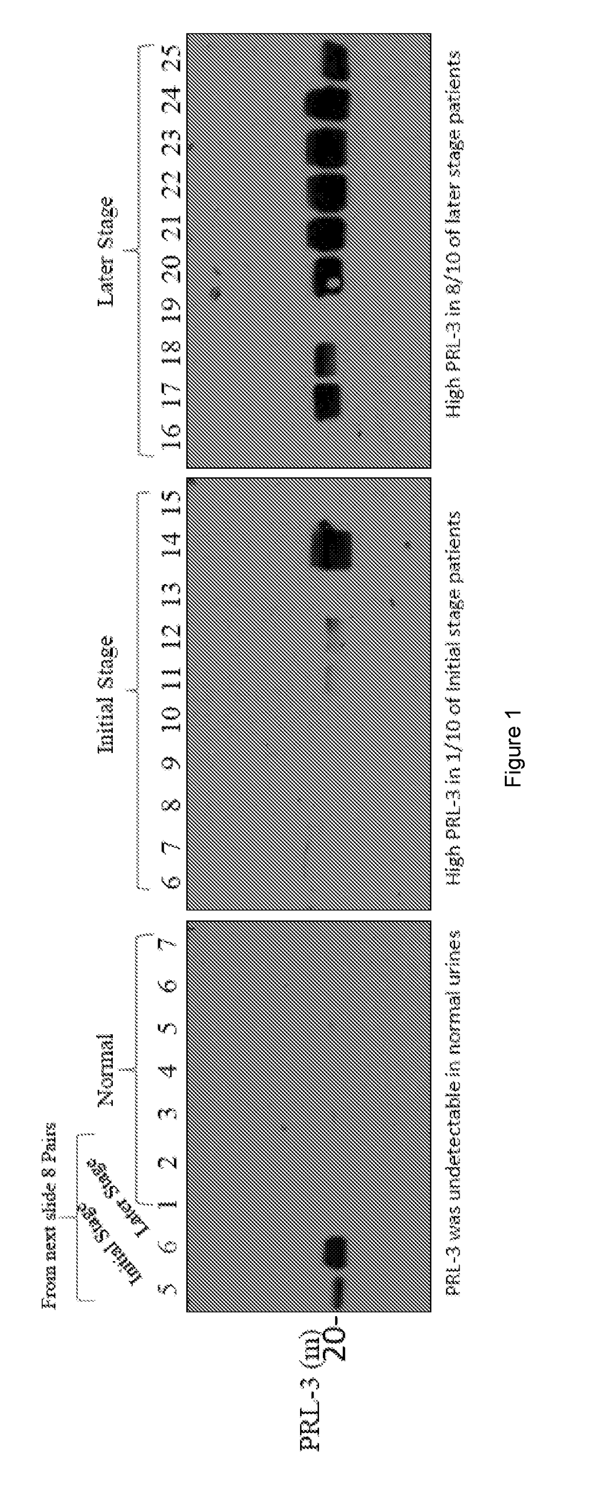
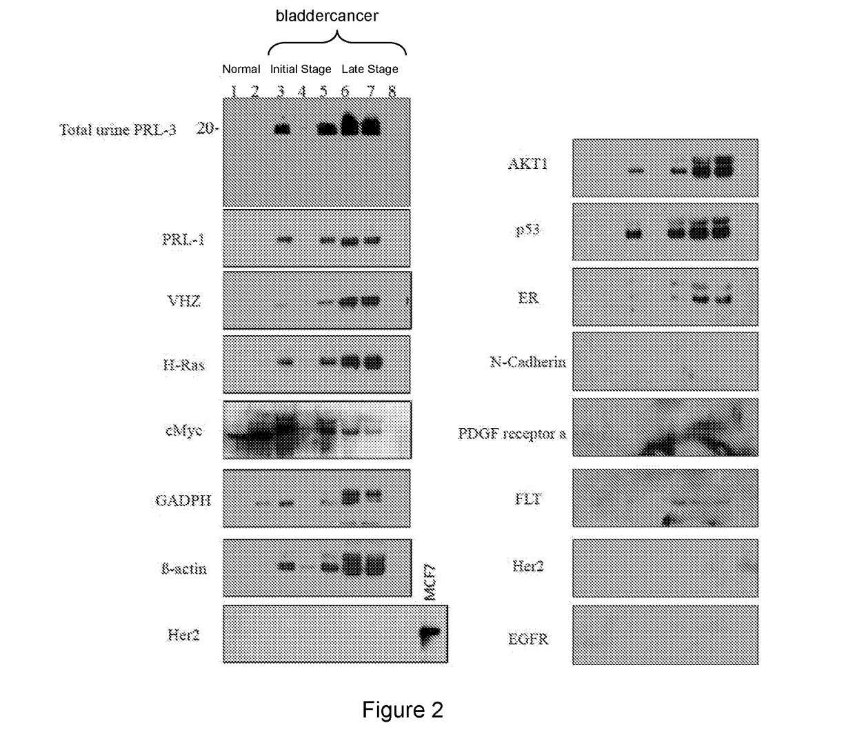
[0240]We examined intracellular oncoproteins: PRL-3, PRL-1, VHZ, c-myc, H-ras, AKT-1, p53, Rac1, FAK, Runx1, Estrogen Receptor, PTEN, b-actin, GAPDH from 101 cancer urines (26 bladders, 44 lungs, 10 breasts, 6 Stomach, 15 NPC), and 11 normal urines. We often detect these intracellular oncoproteins in cancer urines. We also detect mutant forms of GAPDH and b-Actin in cancer (but not normal) urines. The results are show in FIGS. 1-8.

[0241]We examined extracellular oncoproteins: Her2, N-Cadherin, PDGF receptor (Alpha), FLT-3, p-EGFR from 81 cancer urines (6 bladders, 44 lungs, 10 breasts, 6 Stomach, 15 NPC), and 11 normal urines, we only detected FLT-3 at low levels in 3 out of 6 bladder cancer urines using western blot.

[0242]We detected intracellular oncoproteins (much more frequently) than extracellular oncoproteins in cancer urines but not in normal urines. Without wishing to be bound by theory this suggests that extracellular oncoproteins could be more tightly associated with lipid...

example 2

Exosome Isolation and Kits

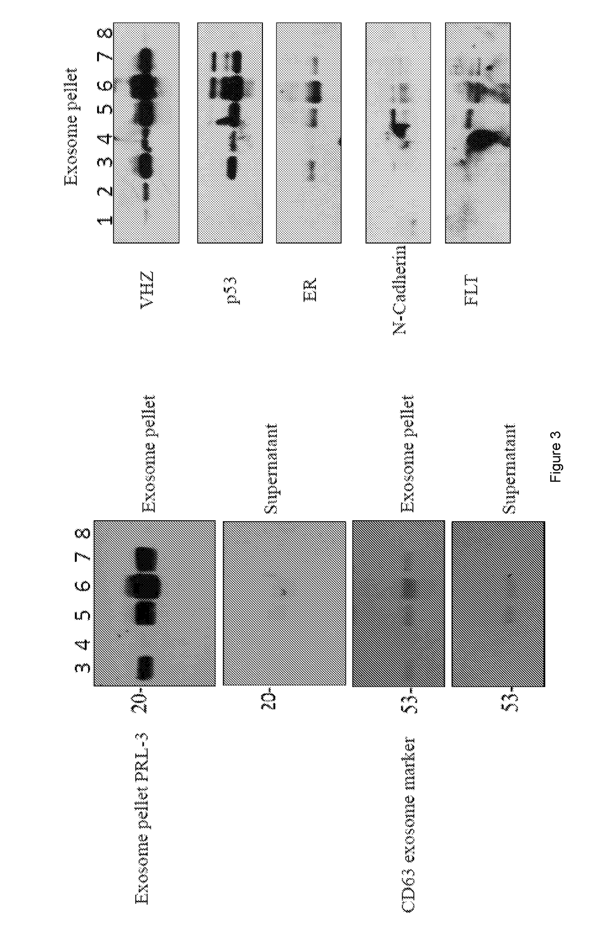
[0243]This example provides a sample protocol for obtaining proteins from exosomes in a bodily fluid sample. The protocol is represented in FIG. 9.

[0244]Protocol: Cat No. EXOAB-KIT-1

[0245]If samples are frozen, thaw on ice

[0246]Combine 10 ml samples+2 ml ExoQuick-TC i.e, (if 500 ul of samples then 100 ul of ExoQuick-TC)

[0247]Mix well by inversion three times

[0248]Place at 4° C. for overnight

[0249]Centrifuge at 1500*g for 30 minutes or (1.5 rfc)

[0250]Remove supernatant, keep Exsosome pellet

[0251]Centrifuge at 1500*g for 5 minutes to remove all trace of fluid

[0252]Add 100 ul RIPA (radio immunoprecipitation assay) buffer or PBS (according to pellet) to exosome pellet and vortex briefly for 15 seconds.

[0253]Place in the room temperature for 20 minutes and take the protein reading at 595 nm.

[0254]Add SDS dye and heat up the samples for 5 minutes at 100° C.

[0255]Perform the Western blot.

example 3

Evaluation of Prototype Rapid Test Device.

[0256]I. Purpose:[0257]1. Study the feasibility to develop a prototype rapid test device for the detection of biomarker PRL3 in patient urine samples.

[0258]II. Background

[0259]Bio-marker PRL3 was found by Dr. Zeng Qi of IMCB of National University of Singapore in 1998. It had been showed its elevated level in thousands of cancer patient. Dr. Zeng had developed two monoclonal and two rabbit antibodies anti-PRL3. This study intends to evaluate these four antibodies and to investigate the feasibility to use these antibodies to develop an immunochromatographic device using lateral flow technology to distinguish the urine between cancer and normal person.

[0260]Rapid test format (lateral flow device) has the advantage of short assay time (only 10-20 minutes to see the test results). Assay also can be performed at the sense or doctor office without special equipment. Dr. Zeng has the interesting to develop such rapid test and contracted AD Consulta...


PUM

PropertyMeasurementUnit
diameteraaaaaaaaaa
solubleaaaaaaaaaa
sizeaaaaaaaaaa

Abstract

The data provided herewith show that, biomarkers found within the cells of a tumor or cancer may also be present at adequate levels within the patient's urine to enable detection. Method of diagnosis, patient selection and treatment are provided, along with kits and devices.

Description

FIELD OF THE INVENTION[0001]The present invention relates to the fields of cell biology, molecular biology and biochemistry. This invention also relates to the field of medicine. In particular, it relates to diagnosis of diseases, in particular cancer.BACKGROUND TO THE INVENTION[0002]Cancer has been the subject of intense investigation in the past two decades. However, the underlying causes responsible for cancer metastasis are poorly understood and most types of cancer prevention are still limited.[0003]Antibody based therapy has proven to be effective for cancer treatment; however, this approach has traditionally been limited to extracellular or secreted proteins expressed by cancer cells. Thus, a number of potential cancer or tumour markers and cancer antigens have been identified in the literature and antibody therapies have been developed against some of them.[0004]For example, the well-known cancer therapy Herceptin (Trastuzumab) is a monoclonal antibody that can kill HER2-pos...

Claims


Application Information

Patent Timeline
Patent Type & Authority Applications(United States)
IPC IPC(8): G01N33/574C07K16/40
CPCG01N33/5748G01N2333/82C12Y301/03048C07K16/40G01N33/57488A61K2039/505G01N2333/916A61P35/00
Inventor ZENG, QI
Owner AGENCY FOR SCI TECH & RES
Why Eureka
Try Eureka