

Method and System for Analyzing Image Data
a technology of image data and analysis method, applied in the field of digital image analysis, can solve the problems of skull stripping, spatial unbiased, image cropping,
- Summary
- Abstract
- Description
- Claims
- Application Information
AI Technical Summary
Benefits of technology
Problems solved by technology
Method used
Image

Examples
Embodiment Construction
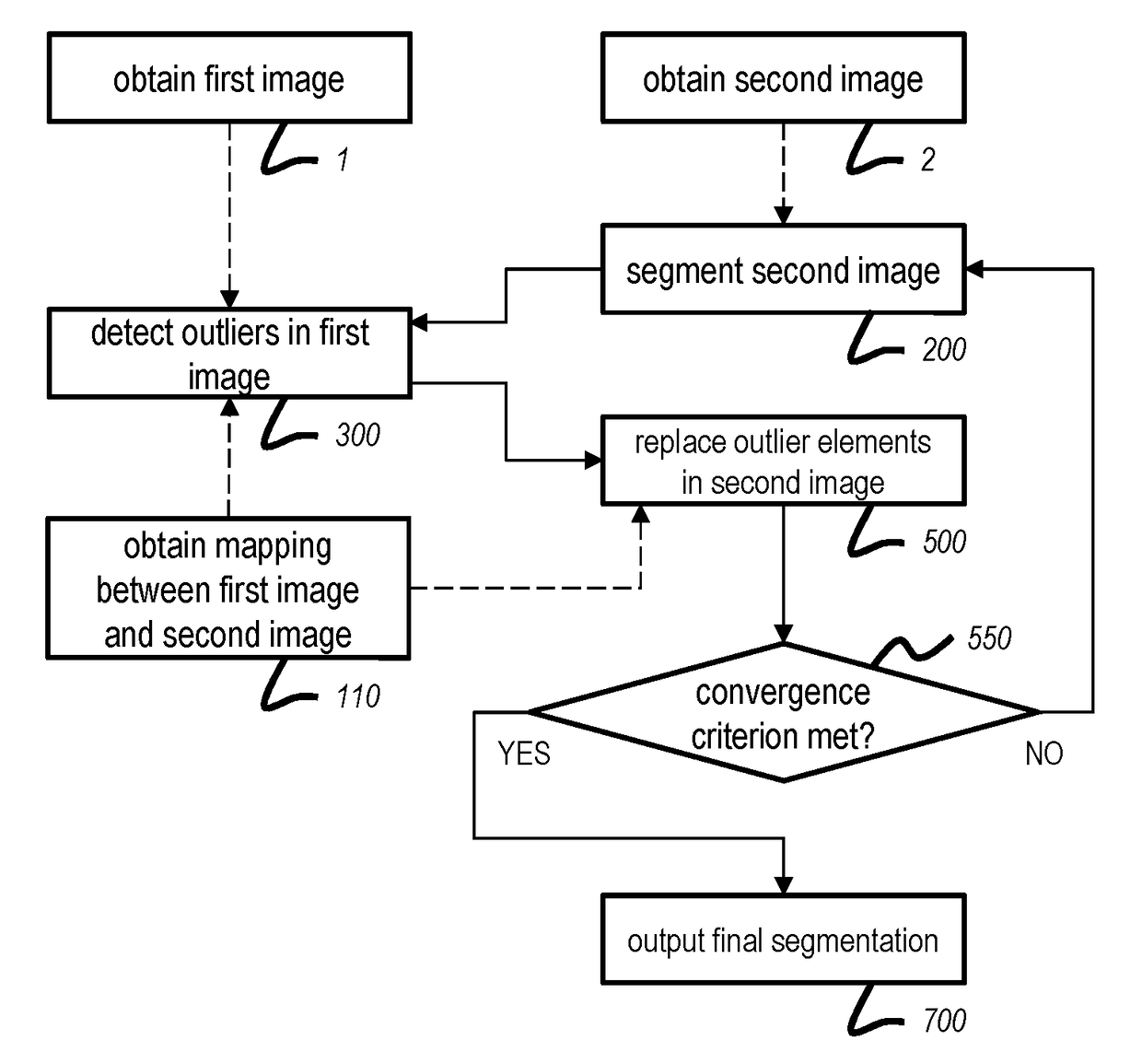
[0038]FIG. 1 provides a flow chart of a method of analyzing image data according to a general embodiment of the present invention.
[0039]The illustrated method comprises obtaining 1 a first image 10 of a first part of an object and obtaining 2 a second image 20 of a second part of the object, the second part having substantial overlap with the first part. The images may be obtained from storage, or directly from an imaging device. The method further comprises obtaining a mapping 110 between the first image 10 and the second image 20. The mapping may be available from storage, or may be produced on the fly by applying a registration algorithm.
[0040]The second image 20 is segmented 200 to obtain a segmentation, i.e. different elements (pixels or voxels) of the second image 20 are classified into various predefined categories. The segmentation information is transferred to the first image 10 on the basis of the aforementioned mapping. In the first image 10, outliers are detected 300 on ...
PUM
 Abstract
Description
Claims
Application Information