
Mobile three-lead cardiac monitoring device and method for automated diagnostics

a cardiac monitoring and three-lead technology, applied in the field of bioelectric signal recording methods and methods, can solve the problems of difficult, unreasonable, and inability to reliably use for a patient to record a full 12l ecg by himself, and achieve the effect of eliminating baseline wandering and/or muscle noise, and avoiding power line interferen

Active Publication Date: 2017-10-12
HEARTBEAM INC
View PDF19 Cites 5 Cited by
  • Summary
  • Abstract
  • Description
  • Claims
  • Application Information

AI Technical Summary

Benefits of technology

The patent text is describing methods for processing two electrical cardiac recordings to eliminate interference, baseline wandering, and muscle noise. These methods also aim to obtain a representative beat using fiducial points and a median beat procedure, and check for switching of the left and right finger. The technical effects of these methods are improved accuracy and reliability in measuring electrical activity of the heart.

Problems solved by technology

However, devices recording only one ECG lead may be used only for rhythm disorders.
Because the ECG changes needed for detection of an AMI may occur in as few as only two among 12 leads of a conventional ECG, it may be difficult, to reliably use only a single lead (or in some cases only a few leads) to reliably and thoroughly detect AMI.
Further, it is also unreasonable for a patient to record a full 12L ECG by himself, because of the difficulty in placing the leads.
All these solutions have significant drawbacks.
For example, all of these solutions typically require complicated measuring procedures (such as with Heartview, Smartheart), and may require attaching electrodes by the means of cables, taking the clothes off from the waist up, using straps and multi-step recording procedure (e.g., see US20120059271A1 to Amitai et al.).
Unfortunately, the heart dipole is only the first term in the multipole mathematical expansion of diffuse heart electrical activity and this approximation is valid only for recording points at a sufficient distance from the heart.
Further the described reconstruction techniques for converting a few leads into a 12 lead signal for analysis by a cardiologist or other technical expert are also limited.
These systems also have additional drawbacks.
It was observed in clinical studies using Cardiobip that breast in female patients and pronounced pectoralis muscle in male patients may prevent a reliable contact of all three electrodes with the chest surface simultaneously.
It has also been observed that the symmetrical arrangement of finger electrodes on the front side of the device may cause switching of left and right hand fingers in about 10% recordings, making the recording useless for diagnostics.
Similarly, other solutions that use a reduced set of three leads (e.g., US20140163349A1; US20100076331) typically use the three leads that are coplanar and therefore lack enough diagnostic information for AMI detection.
In addition, the requirement for trained medical personnel for the interpretation of recorded ECG may be an organizational challenge and increases the operational cost of the system, and the accuracy of the human ECG interpretation may have large variance.
Automated software for ECG interpretation is also used in the systems for early diagnosis of AMI, but they have performance that is inferior to that of human interpreters.
Unfortunately, a large number of patients (up to 15%) presenting with chest pain have STE of non-ischemic etiology (NISTE) on their presenting (to the emergency room) ECG.
Thus, both human readers and automated software may often misinterpret NISTE as a new STE due to ischemia.

Method used


Image

  • Mobile three-lead cardiac monitoring device and method for automated diagnostics
  • Mobile three-lead cardiac monitoring device and method for automated diagnostics
  • Mobile three-lead cardiac monitoring device and method for automated diagnostics

Examples


examples

[0127]A clinical study was done to evaluate the diagnostic accuracy of the methods described above for detecting myocardial ischemia provoked by balloon inflation in coronary arteries during a PCI (Percutaneous Coronary Intervention) procedure.

[0128]In this example, data was acquired continuously. Continuous data from standard 12 lead ECG with additional three special leads were obtained from each patient during the entire period of balloon occlusion, and for a short period before and after. Target duration for balloon occlusion was at least 90 sec if the patient is stable. In each patient, one baseline recording was taken prior to the beginning of the PCI procedure, and one pre-inflation during the procedure, prior to first balloon insertion. In each lesion / intervention site, one inflation recording was taken just before the balloon deflation. The analyzed data set contains ECG recordings of 66 patients and 120 balloon occlusions (up to three arteries inflated per patient).

[0129]Da...


PUM


Abstract

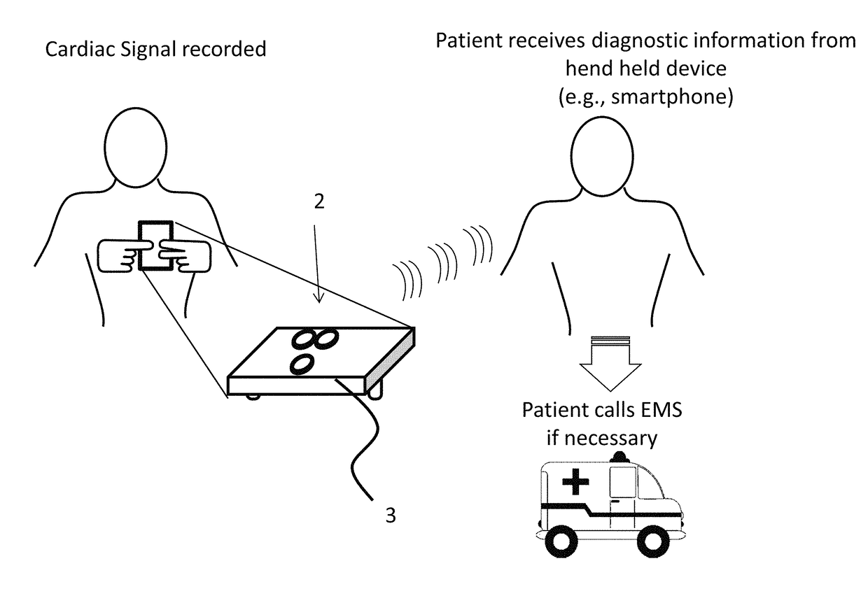
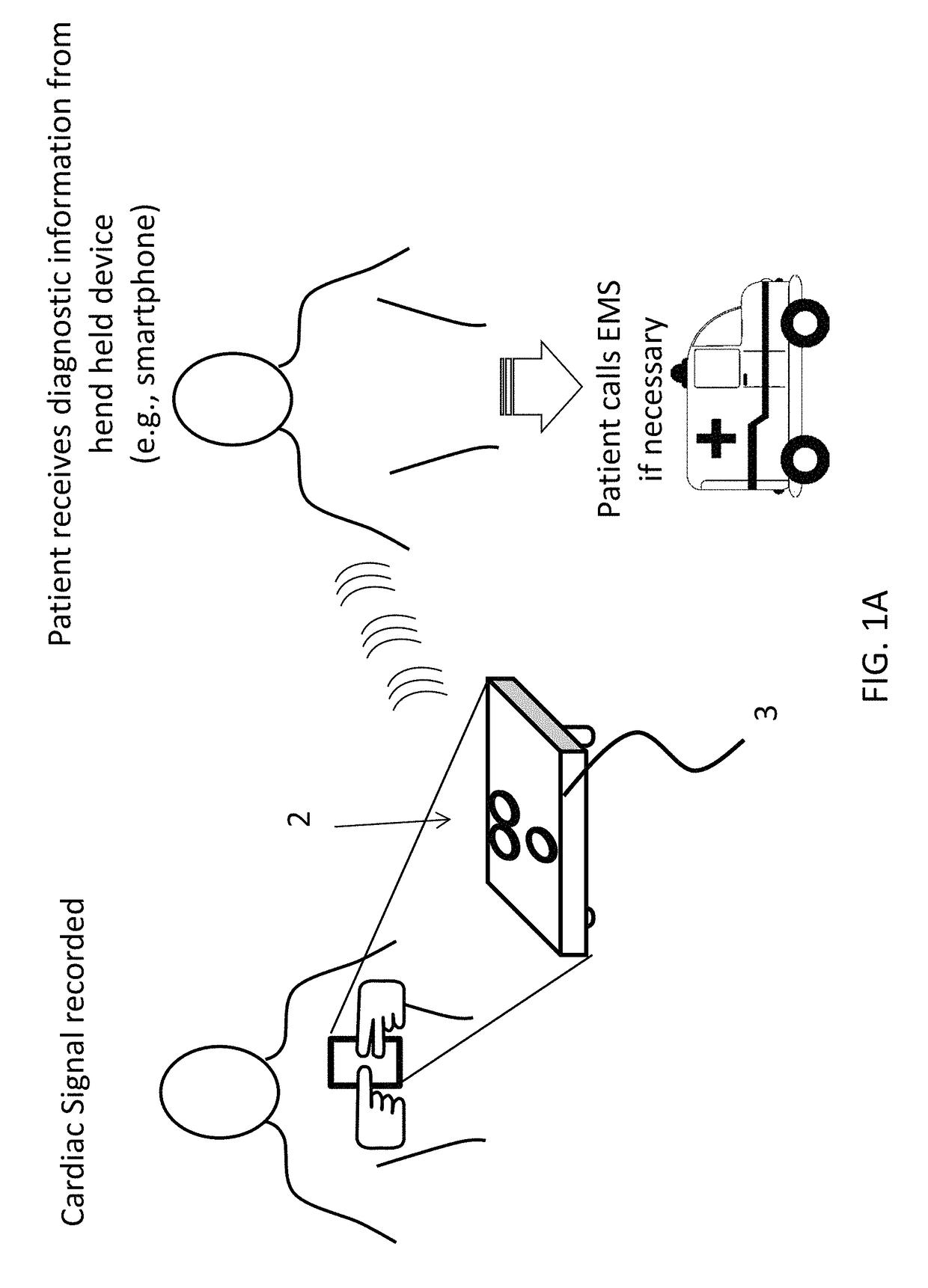
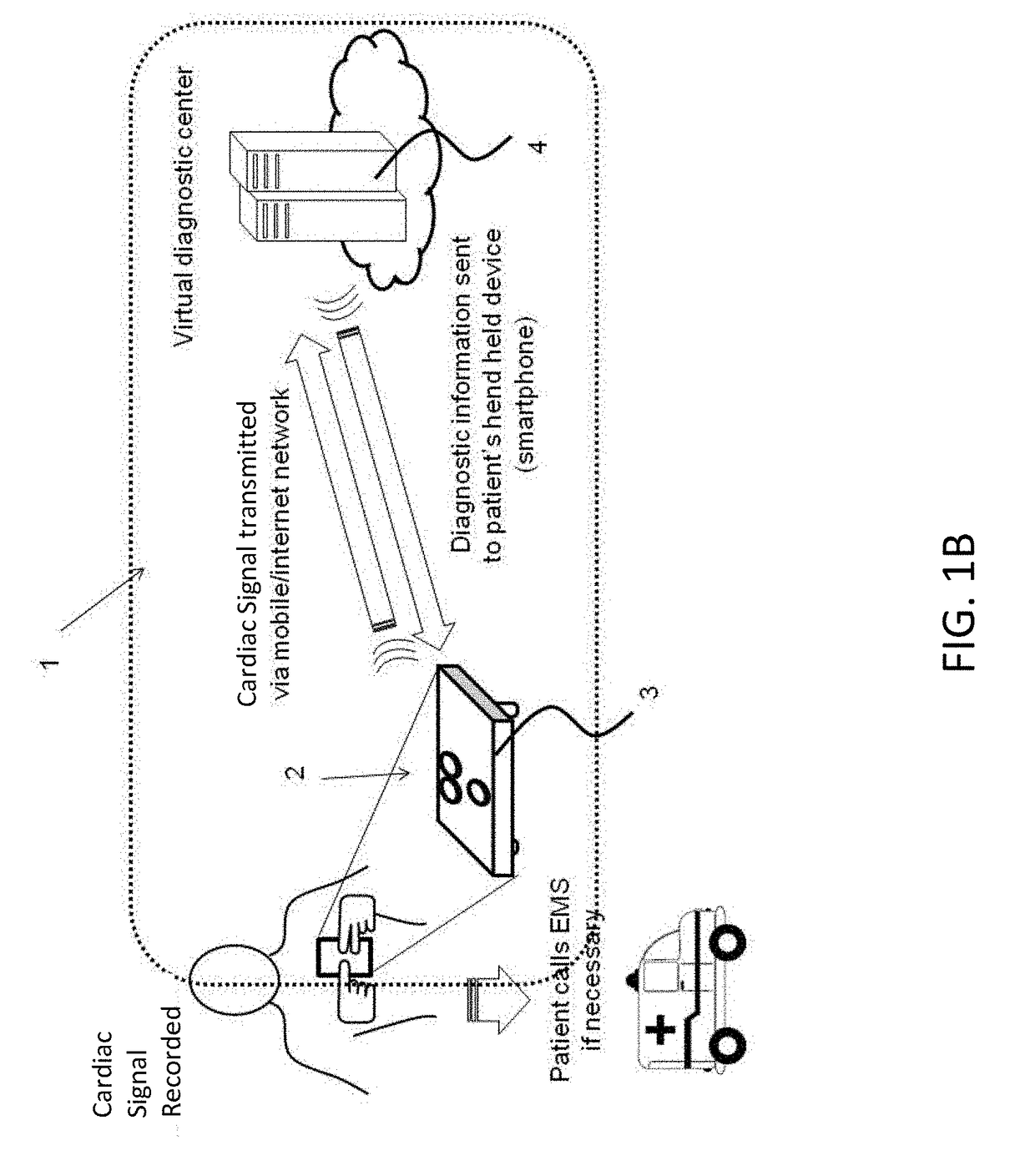
Methods and apparatuses, including devices and systems, for remote and detection and / or diagnosis of acute myocardial infarction (AMI). In particular, described herein are handheld devices having an electrode configuration capable of recording three orthogonal ECG lead signals in an orientation-specific manner, and transmitting these signals to a processor. The processor may be remote or local, and it may automatically or semi-automatically detect AMI, atrial fibrillation or other heart disorders based on the analyses of the deviation of the recorded 3 cardiac signals with respect to previously stored baseline recordings.

Description

CROSS REFERENCE TO RELATED APPLICATIONS[0001]This application is a continuation of U.S. patent application Ser. No. 15 / 096,159, filed Apr. 11, 2016, titled “MOBILE THREE-LEAD CARDIAC MONITORING DEVICE AND METHOD FOR AUTOMATED DIAGNOSTICS,” which claims priority to U.S. Provisional Patent Application No. 62 / 145,431, filed Apr. 9, 2015, titled “MOBILE THREE-LEAD CARDIAC MONITORING DEVICE AND METHOD FOR AUTOMATED DIAGNOSTICS”, both of which are incorporated herein by reference in their entirety.INCORPORATION BY REFERENCE[0002]All publications and patent applications mentioned in this specification are herein incorporated by reference in their entirety to the same extent as if each individual publication or patent application was specifically and individually indicated to be incorporated by reference.FIELD[0003]Described herein are methods and apparatuses for recording bioelectric signals. More particularly, described herein are hand held apparatuses (devices and systems) for recording,...

Claims


Application Information

Patent Timeline
Patent Type & Authority Applications(United States)
IPC IPC(8): A61B5/0404A61B5/046A61B5/04A61B5/0408A61B5/00A61B5/332A61B5/361G16Z99/00
CPCA61B5/0404A61B5/04085A61B5/0006A61B5/04012A61B5/046G06F19/345A61B5/747A61B5/684A61B2560/0468A61B2562/0209G06F19/3418A61B5/6898G16H40/63G16H50/20A61B5/6823A61B5/6825G16H40/67A61B5/341A61B5/332A61B5/316A61B5/282A61B5/361G16Z99/00A61B5/346A61B5/33
Inventor BOJOVIC, BOSKOHADZIEVSKI, LJUPCOVUKCEVIC, VLADANMITROVIC, UROSMILETIC, MARJAN
Owner HEARTBEAM INC
Learn More