
Model-based segmentation of an anatomical structure

a model-based segmentation and anatomical technology, applied in the field of model-based segmentation of anatomical structures, can solve the problem that the imaging modality used in acquiring medical images may only provide a limited field of view of the patient's interior, and achieve the effect of facilitating the further processing of the adapted model

Active Publication Date: 2017-11-23
KONINKLJIJKE PHILIPS NV
View PDF13 Cites 2 Cited by
  • Summary
  • Abstract
  • Description
  • Claims
  • Application Information

AI Technical Summary

Benefits of technology

The patent text is about a system or method that can adapt a model to a medical image that only shows part of a patient's anatomical structure. This adapted model can then be further processed. The text also describes how the system can use a regularization term to make the transformation estimation more stable when there is less anatomical structure shown in the image. The text also talks about a method for performing extrapolation that reduces internal energy by minimizing the relationship between vertices and / or edges of the model. Overall, the technical effects of this patent text are improved accuracy and stability in adapting models to medical images and facilitating further processing of the adapted models.

Problems solved by technology

A reason for this may be that the imaging modality used in acquiring the medical image may only provide a limited field of view of the patient's interior.

Method used


Image

  • Model-based segmentation of an anatomical structure
  • Model-based segmentation of an anatomical structure
  • Model-based segmentation of an anatomical structure

Examples


Embodiment Construction

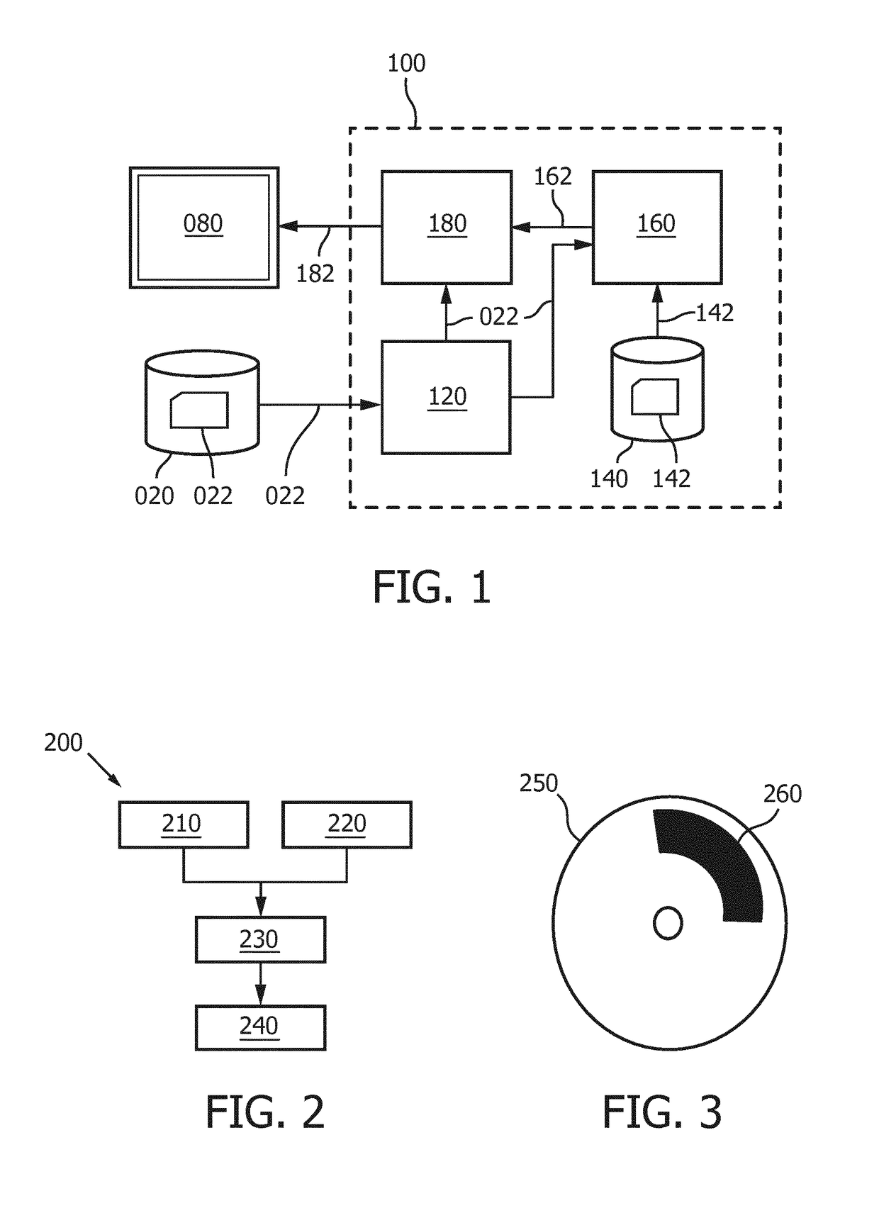
[0078]FIG. 1 shows a system 100 for model-based segmentation. The system 100 comprises an image interface 120 for accessing image data 022 representing a medical image of a patient. In the example of FIG. 1, the image interface 120 is shown to be connected to an external image repository 020. For example, the image repository 020 may be constituted or be part of a Picture Archiving and Communication System (PACS) of a Hospital Information System (HIS) to which the system 100 may be connected or comprised in. Accordingly, the system 100 may obtain access to the medical image 022. In general, the image interface 120 may take various forms, such as a network interface to a local or wide area network, such as the Internet, a storage interface to an internal or external data storage, etc.

[0079]It is noted that throughout this text and where appropriate, a reference to the medical image is to be understood as a reference to the medical image's image data.

[0080]The system 100 further compr...


PUM


Abstract

A system and method is provided for performing a model-based segmentation of a medical image which only partially shows an anatomical structure. In accordance therewith, a model is applied to the image data of the medical image, the model-based segmentation providing an adapted model having a first model part having been adapted to the first part of the anatomical structure in the medical image of the patient, and a second model part representing the second part of the anatomical structure not having been adapted to a corresponding part of the medical image. Metadata is generated which identifies the first model part to enable the first model part to be distinguished from the second model part in a further processing of the adapted model. Advantageously, the metadata can be used to generate an output image which visually indicates to the user which part of the model has been personalized and which part of the model has not been personalized. Other advantageous uses of the metadata in the further processing of the adapted model have also been conceived.

Description

FIELD OF THE INVENTION[0001]The invention relates to a system and a method for performing a model-based segmentation of an anatomical structure in a medical image. The invention further relates to a workstation and imaging apparatus comprising the system and to a computer program product comprising instructions for causing a processor system to perform the method.BACKGROUND OF THE INVENTION[0002]Robust automatic segmentation of various anatomical structures in a medical image is a key enabler in improving clinical workflows. Here, the term segmentation refers to the identification of the anatomical structure in the medical image by, e.g., delineation of the boundaries of the anatomical structure, by labeling of the voxels enclosed by the boundaries, etc. Once such segmentation has been performed, it is possible to extract clinical parameters such as, in case of, e.g., a cardiac structure, ventricular mass, ejection fraction and wall thickness. Additionally or alternatively, the segm...

Claims


Application Information

Patent Timeline
Patent Type & Authority Applications(United States)
IPC IPC(8): G06T7/00G06T7/149
CPCG06T7/0012G06T7/149G06T2207/30048G06T2207/10132G06T2207/10116G06T2207/20128
Inventor WEBER, FRANK MICHAELBUERGER, CHRISTIANNIJHOF, NIELSMAZOUER, PHILIPPE PAULWAECHTER-STEHLE, IRINA
Owner KONINKLJIJKE PHILIPS NV
Learn More