Stain-free histopathology by chemical imaging

Active Publication Date: 2017-12-14
THE BOARD OF TRUSTEES OF THE UNIV OF ILLINOIS
View PDF6 Cites 1 Cited by
  • Summary
  • Abstract
  • Description
  • Claims
  • Application Information

AI Technical Summary

Benefits of technology

[0005]Provided herein are methods and systems for generating or obtaining an image of a sample, such as biopsy tissue sections, without the use of dyes or stains (such as those currently used in histopathology and IHC). Spectroscopic imaging (such as Fourier transform infrared (FT-IR) spectroscopic imaging) is used to record the intrinsic chemical composition of sample, and numerical algorithms are employed to relate the biochemical content of the IR spectral data to structure of the sample, such as tissue s

Problems solved by technology

Unfortunately, tissues have little contrast in brightfield optical imaging (FIG. 1A), and require the use of stains or dyes to provide contrast.
While advances have been made (Ruifrok et al., Anal Quant Cytol Histol 23:291-9,2001; Taylor et al., Histopathology 49

Method used


Image

  • Stain-free histopathology by chemical imaging
  • Stain-free histopathology by chemical imaging
  • Stain-free histopathology by chemical imaging

Examples


Example

EXAMPLE 1

Materials and Methods

[0118]This example provides the materials and methods used in Examples 2-4 below. Although the examples describe studies using breast tissue, one skilled in the art will appreciate that other tissues and samples can be used based on the teachings provided herein.

[0119]The problem of spectral unmixing in breast tissue was solved herein by mapping the infrared images to bright-field images representing an array of common histological stains. Several tissue samples representing a broad panel of normal tissue, non-malignant and cancer subtypes, including tissue microarrays and surgical resections of breast biopsies, were used. A training set was constructed by first imaging the tissue using mid-infrared spectroscopy. Adjacent sections were then stained using a panel of standard and immunohistochemical stains and imaged using bright-field microscopy. The bright-field images were then adjusted to overlay the spectroscopic images. This created a spatial (pixel...

Example

EXAMPLE 2

Comparison of H&E Stained Image to Computed Stain Image

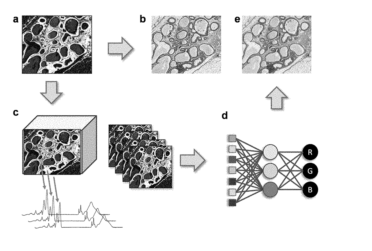
[0136]Breast tissue previously fixed and paraffin embedded, was stained with H&E, and imaged using microscopy before and after staining with H&E. As shown in FIG. 1A, brightfield optical image of tissue has little contrast. Conventionally, the use of H&E stain allows a visualization of tissue morphology using optical microscopy (FIG. 1B).

[0137]The same unstained tissue shown in FIG. 1A was subjected to IR spectroscopic imaging in the form of two spatial dimensions and a spectral dimension (FIG. 1C). The spectrum at every pixel contains specific features that are indicative of the molecular content of the sample as well as its optical properties (Davis et al., Anal. Chem. 82:3474-86, 2010).

[0138]The size of the data is then reduced from either knowledge of biochemical absorption or a statistically-based dimensionality reduction approaches. This produces a data set containing a significantly smaller number of spectral fea...

Example

EXAMPLE 3

Comparison of Tissue-Specific Stained Images to Computed Stain Images

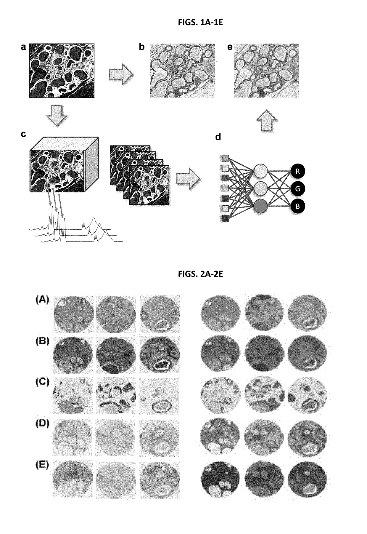
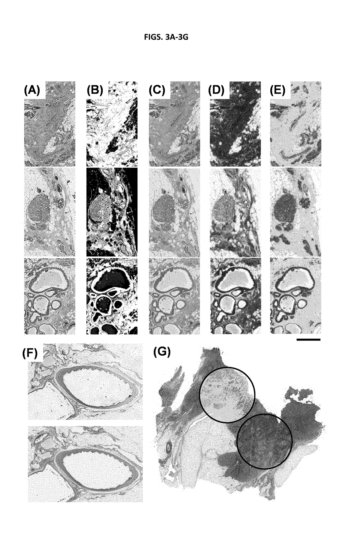
[0141]H&E stains are fairly non-specific in terms of the functional or molecular content of the tissue. Visualizing molecular content is significantly more expensive, time-consuming and difficult; yet, this is precisely the origin of the contrast mechanism in chemical imaging. Hence, additional stains were examined that are indicative of tissue function and integrity. Immunohistochemical (IHC) stains for cell types are often used in diagnostic imaging or for specific research purposes, for example, high molecular weight (HMW) cytokeratin (epithelial-type cells), vimentin (fibroblast-like cells), smooth muscle alpha actin (myo-like cells), P63 (myoepithelial cells), CD31 (endothelial cells) and Masson's Trichrome stain (collagen and keratin fibers), are commonly employed.

[0142]A comparison of the physically stained and computationally stained images is shown in FIGS. 2A-2E. Since the process is correlative,...


PUM


Abstract

The present disclosure provides methods, systems, and computer-readable storage media that can be used to image an unstained sample. Traditionally histopathology and immunohistochemistry methods use stains or dyes in combination with microscopy (or other detection methods) to detect cells and cellular structures, such as proteins. However, the disclosed methods do not require the use of such stains and dyes. The disclosed methods can include obtaining a spectroscopic image (e.g., infrared (IR) imaging data) of the sample, analyzing the resulting spectroscopic image to reduce the dimensionality of the spectroscopic image, comparing the reduced spectroscopic image compared to a control (e.g., by using an appropriately trained algorithm) and generating an output computed stain image from the reduced IR spectra, thereby imaging the sample without the use of stains or dyes.

Description

CROSS REFERENCE TO RELATED APPLICATION[0001]This application is a continuation of and claims priority to U.S. patent application Ser. No. 14 / 211,292 filed Mar. 14, 2014, which claims the benefit of U.S. Provisional Application No. 61 / 794,171, filed Mar. 15, 2013. The contents of each of the foregoing are hereby incorporated by reference into this application as if set forth herein in full.ACKNOWLEDGMENT OF GOVERNMENT SUPPORT[0002]This invention was made with government support under 1R01EB009745 awarded by The National Institutes of Health. The government has certain rights in the invention.FIELD[0003]The disclosure provides methods of imaging biological samples to provide a molecular composition of the sample without the use of stains.BACKGROUND[0004]Clinical decisions and biomedical research rely significantly on imaging the architecture and morphology of tissues. Unfortunately, tissues have little contrast in brightfield optical imaging (FIG. 1A), and require the use of stains or...

Claims


Application Information

Patent Timeline
IPC IPC(8): G06T7/00G06K9/00
CPCG06T2207/30024G06T2207/20084G06T2207/20081G06T2207/10056G06T2207/10048G06T2207/10024G06K9/00127G06T7/0014G06K9/0014G06V20/69G06V20/695
Inventor BHARGAVA, ROHIT
Owner THE BOARD OF TRUSTEES OF THE UNIV OF ILLINOIS
Why Eureka
Try Eureka