Method and system for detecting pathological anomalies in a digital pathology image and method for annotating a tissue slide

a pathology image and digital technology, applied in image enhancement, image analysis, instruments, etc., can solve the problems of human error risk, high visual and subjective process of annotating tissue slides, and the need for a trained specialis

Active Publication Date: 2017-12-28
INIFY LAB AB
View PDF6 Cites 23 Cited by
  • Summary
  • Abstract
  • Description
  • Claims
  • Application Information

AI Technical Summary

Benefits of technology

[0005]The subject of the present invention is to

Problems solved by technology

Thus, interpreting and annotating tissue slides is a highly visual and subjective process which is time consuming, costly, require a trained specialist, and involves the risk for human errors.
For ex

Method used


Image

  • Method and system for detecting pathological anomalies in a digital pathology image and method for annotating a tissue slide
  • Method and system for detecting pathological anomalies in a digital pathology image and method for annotating a tissue slide
  • Method and system for detecting pathological anomalies in a digital pathology image and method for annotating a tissue slide

Examples


Embodiment Construction

[0051]In the following a number of embodiments of the invention are shown and described. The same reference numbers have been used for the same or similar features throughout the description in the embodiments disclosed below.

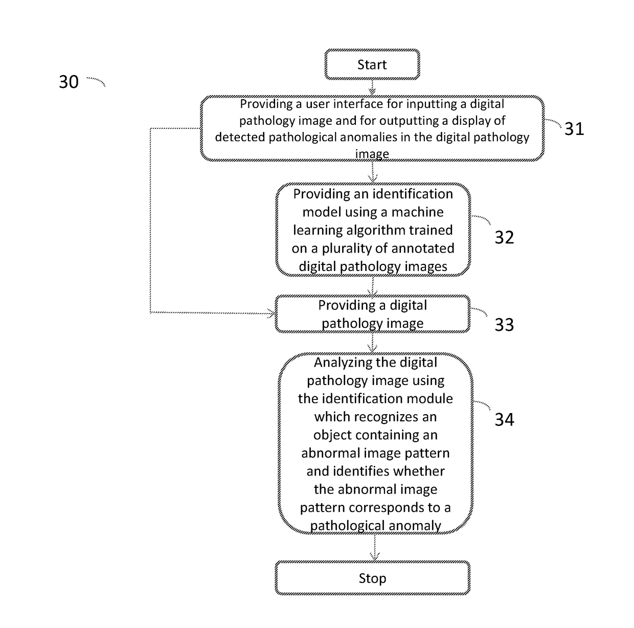
[0052]FIG. 1 is a flow diagram illustrating the steps of a method 10 performed by a computing system 50, 50′, 50″, 50′″ (the system is described further down in the text; in the text describing FIG. 5-7) for detecting pathological anomalies in a digital pathology image, i.e. a visual representation of the interior of a body for clinical analysis, according to an embodiment of the present invention. Said pathological anomaly can for example be an infection or an inflammation or a cancer tumour. The infection can for example be cholera, dengue or malaria and the inflammation can for example be appendicitis, bursitis or colitis. The cancer tumour can for example be a prostate cancer tumour or a breast cancer tumour.

[0053]The method comprises providing a digital pa...


PUM


Abstract

A method for annotating a tissue slide image, a system and a method performed by a computing system for detecting pathological anomalies in a digital pathology image are disclosed. The method performed by a computing system for detecting pathological anomalies in a digital pathology image includes providing a digital pathology image to the computing system and analyzing the digital pathology image using an identification module arranged on the computing system. The identification module uses a machine learning module to execute recognizing an object containing an abnormal image pattern using an identification model loaded in said identification module and identifying whether the abnormal image pattern corresponds to a pathological anomaly using the identification model.

Description

TECHNICAL FIELD[0001]The present invention relates to a method performed by a computing system for detecting pathological anomalies in a digital pathology image and a system for detecting pathological anomalies in a digital pathology image. The present invention also relates to a method for annotating a tissue slide image.BACKGROUND OF THE INVENTION[0002]A common method to detect pathological anomalies, for example infections, inflammations or cancer tumours, is to acquire a tissue sample by using biopsy during which a small sample of tissue is removed from a part of the body. Tissue samples can also come from a resected tissue / tumour to verify or complement the initial diagnose. The removed tissue sample is then processed and cut into thin sections which are stained to visualize structural features of the tissue sample. The most commonly used stain for this purpose is H&E, which is a combination of the two compounds haematoxylin and eosin. An image of the stained tissue sample is t...

Claims


Application Information

Patent Timeline
IPC IPC(8): G06T7/00G06K9/62
CPCG06T7/0012G06K9/628G06K9/6256G06T2207/20132G06T2207/30024G06T2200/24G06T2207/30096G06K2209/05G06V20/698G06V2201/03G06F18/214G06F18/2431
Inventor EUR N, KRISTIAN
Owner INIFY LAB AB
Why Eureka
Try Eureka