
Handheld device and multimodal contrast agent for early detection of human disease

a multi-modal contrast agent and hand-held technology, applied in the field of hand-held devices and multi-modal contrast agents for early detection of human diseases, can solve the problems of nir radiation from both the external source and the fluorescence of the dye, the obscuration of the wound dressing, and the inability to detect the disease at the same time, so as to improve the accuracy of deep tissue imaging and facilitate diagnosis. rapid

Inactive Publication Date: 2018-03-08
PENN STATE RES FOUND
View PDF0 Cites 3 Cited by
  • Summary
  • Abstract
  • Description
  • Claims
  • Application Information

AI Technical Summary

Benefits of technology

This patent describes a new technology for better imaging deep tissues in the body using nanocolloids that encapsulate both NIR and CT / MRI contrast agents. The nanoparticles are made using calcium phosphosilicate nanoparticles, indocyanine green, and nanoscale magnetite. This technology offers advantages over existing imaging methods like ultrasound and CT, including faster use, lower cost, and wider availability. The nanoparticles can be used as a food additive or tablet, meaning they can be present in a person's body without needing additional administration. Overall, this new technology offers a noninvasive and effective way to image the body's internal tissues.

Problems solved by technology

While the ultrasound systems have permitted rapid diagnosis, issues with the portability (the system is about the size of a larger suitcase), detection limits, operator dependence, obscuration by wound dressings, and lateral resolution remain issues that require improvement.
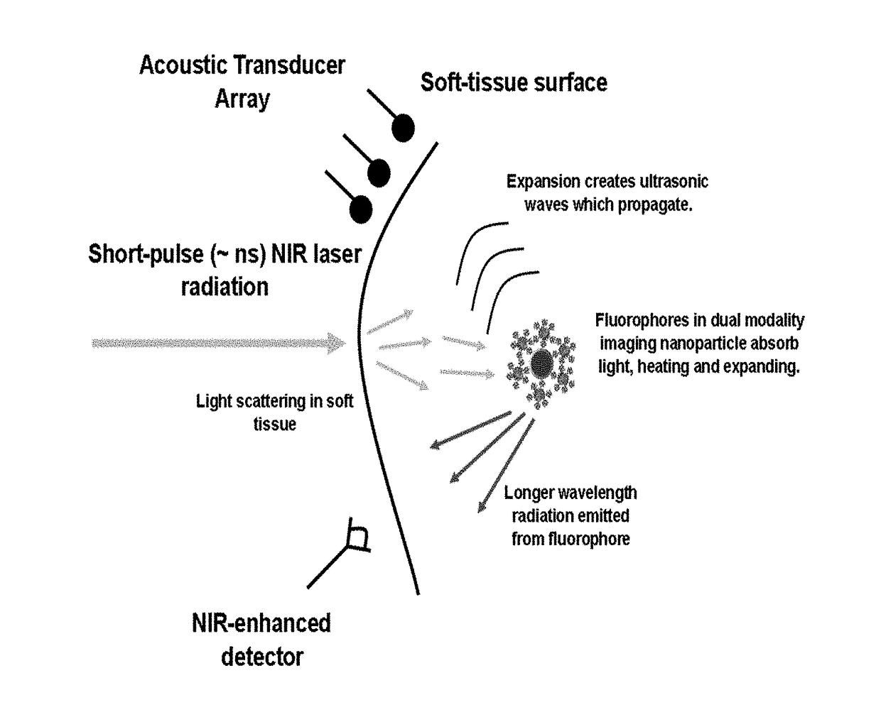
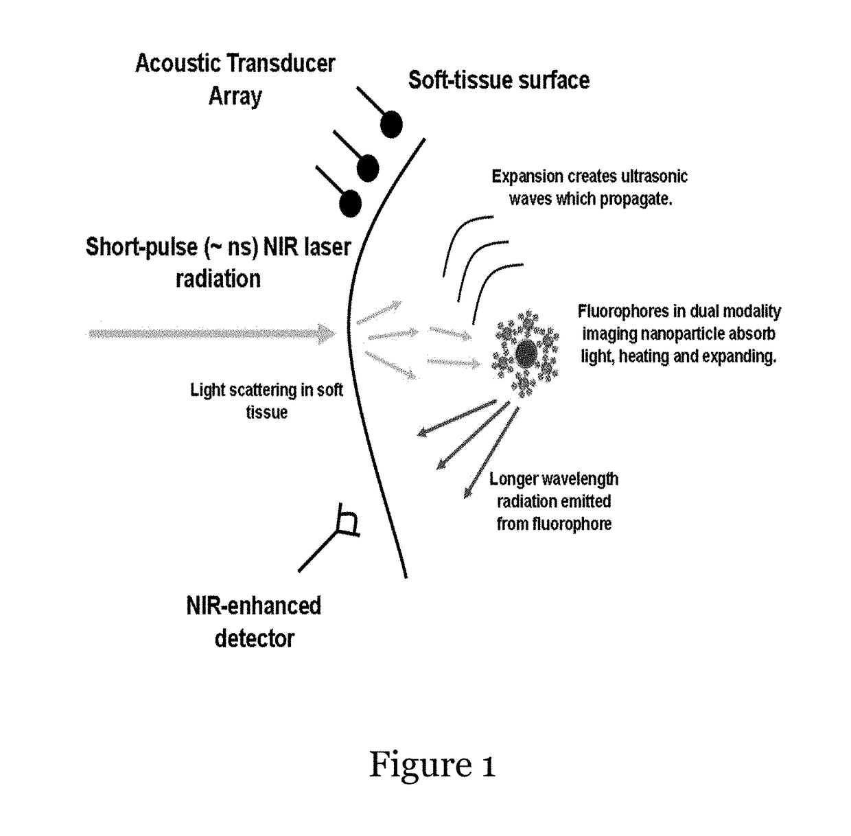
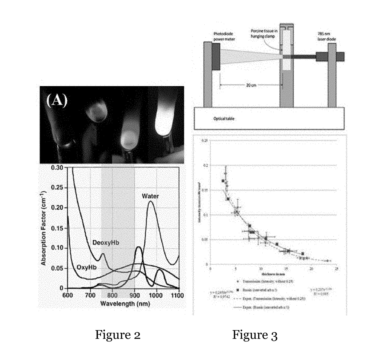
Nevertheless, the NIR radiation from both the external source and the fluorescence of the dye are subject to attenuation, primarily due to scattering events.
Thus, imaging in deeper regions of the body is limited by the power of the external source, the number of fluorescing particles, the quantum efficiency of the fluorophores (number of fluorescence photons per incident photon) and the sensitivity of the detector.
The rapid diagnosis of internal hemorrhage due to blast, crush or blunt trauma is still severely limited by the sensitivity of conventional handheld ultrasound or near infrared imaging devices.

Method used


Image

  • Handheld device and multimodal contrast agent for early detection of human disease
  • Handheld device and multimodal contrast agent for early detection of human disease
  • Handheld device and multimodal contrast agent for early detection of human disease

Examples


example 1

[0086]The magnetite particles of the present disclosure in FIG. 6 have been evaluated as a function of dispersion in MRI and are shown in FIG. 7. While the poorly dispersed magnetite nanoparticles show local hypointense signal areas within each image, the well dispersed magnetite nano-particles produce a uniform signal change over the whole image. It is of great importance to use well dispersed magnetite nanoparticles to be able to draw reliable conclusions about the local concentrations of these particles (e.g. in tissue). Using the poorly dispersed magnetite nanoparticles could over / under estimate the local concentrations. The phantom experiments showed a huge drop of the apparent T2 relaxation time from 96 ms in the Agar phantom without magnetite nanoparticles, to 14 ms with only 0.01 mg / ml magnetite nanoparticles. A further decrease to 1.6 ms was observed when 0.1 mg / ml was used. Acquired T2 maps showed very homogeneous distributions of T2 over the whole phantoms.


PUM


Abstract

Systems comprising a combination of the handheld imaging system with a nanoparticle multimodal contrast agent are disclosed. The imaging system exploits the advantages of both near-infrared emission and the photoacoustic effect by employing calcium phosphosilicate nanocolloid that encapsulates NIR and CT / MRI contrast agents for enhanced deep tissue imaging as well as a portable NIR / PA system using a tunable pulsed laser, CCD imaging technology and acoustic transducer arrays. Methods for using the system, for example in rapid diagnosis of trauma such as that inflicted on a battlefield, are provided.

Description

CROSS-REFERENCE TO RELATED APPLICATIONS[0001]This application claims priority and is related to U.S. Provisional Application Ser. No. 62 / 384,849 filed on Sep. 8, 2016 and entitled “HANDHELD DEVICE AND MULTIMODAL CONTRAST AGENT FOR EARLY DETECTION OF HUMAN DISEASE.” The entire contents of this patent application are hereby expressly incorporated herein by reference including, without limitation, the specification, claims, and abstract, as well as any figures, tables, or drawings thereof.GRANT INFORMATION[0002]This invention was made with government support under Grant No. CA167535, awarded by the National Institutes of Health. The Government has certain rights in the invention.FIELD OF THE DISCLOSURE[0003]The disclosure relates to systems comprising a combination of a handheld imaging system with a nanoparticle multimodal contrast agent. The imaging system exploits the advantages of both near-infrared emission and the photoacoustic effect by employing calcium phosphosilicate nanocoll...

Claims


Application Information

Patent Timeline
Patent Type & Authority Applications(United States)
IPC IPC(8): A61B5/00A61K49/22
CPCA61B5/0095A61K49/222A61K49/221A61B2503/40A61B2560/0431A61B5/0035A61K49/225
Inventor ADAIR, JAMES H.KNECHT, SEAN D.BOYER, J. ERICTUTWILER, RICHARD L.CARR, CONNORTANG, XIAOMENGADAIR, BERNADETTE M.NEUBERGER, THOMASLOC, WELLEY S.WILCZYNSKI, ZACHARY R.MCGOVERN, CHRISTOPHERMATTERS, GAIL L.CHENG, KEITHKESTER, MARKSINOWAY, LAWRENCE
Owner PENN STATE RES FOUND
Learn More