
Charged particle cancer therapy beam state determination system and method of use thereof

a charge particle and beam technology, applied in the field of cancer therapy treatment apparatus, can solve the problems of reducing the ability to repair damaged dna, affecting the treatment effect, and being particularly vulnerable to attack on dna

Active Publication Date: 2018-03-22
PROTOM INT HLDG CORP
View PDF14 Cites 10 Cited by
  • Summary
  • Abstract
  • Description
  • Claims
  • Application Information

AI Technical Summary

Benefits of technology

The patent describes a system for measuring the time it takes for charged particles to travel through a patient's body in a cancer therapy system. The system can help with imaging and treating tumors.

Problems solved by technology

These particles damage the DNA of cells, ultimately causing their death.
Cancerous cells, because of their high rate of division and their reduced ability to repair damaged DNA, are particularly vulnerable to attack on their DNA.

Method used


Image

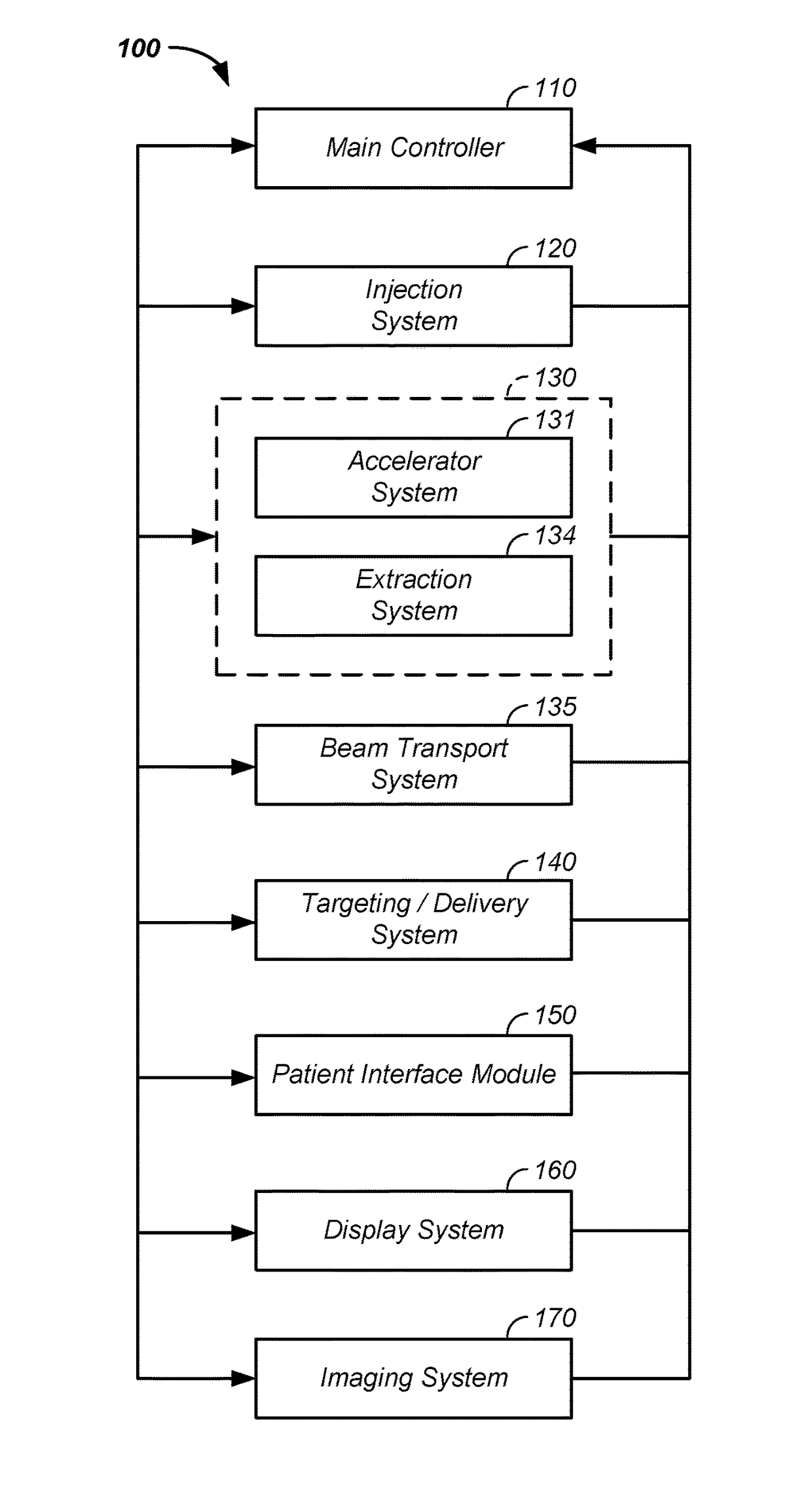
  • Charged particle cancer therapy beam state determination system and method of use thereof
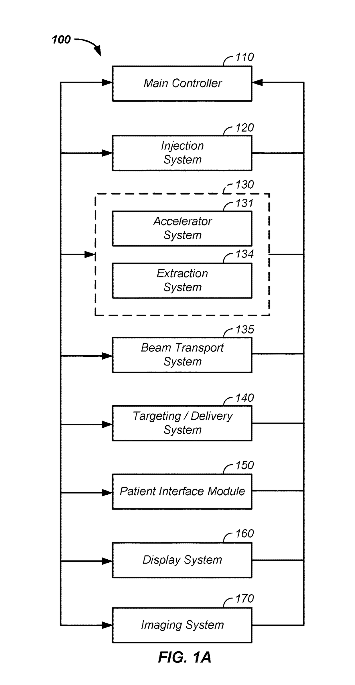
  • Charged particle cancer therapy beam state determination system and method of use thereof
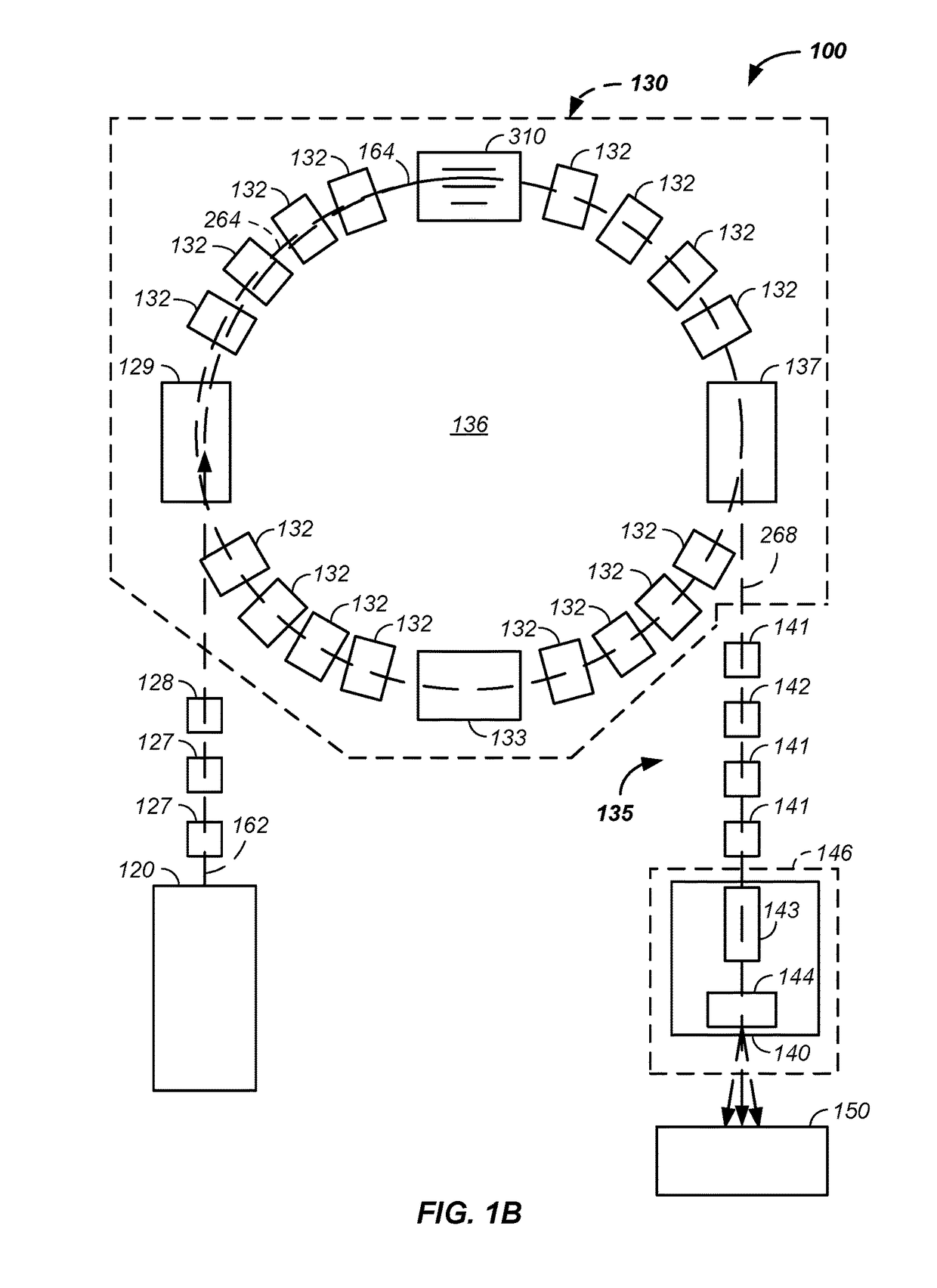
  • Charged particle cancer therapy beam state determination system and method of use thereof

Examples


example vii

[0258]In a seventh example, the rolling floor 1320 forms a continuous loop in the cantilevered three hundred sixty degree rotatable gantry system.

example viii

[0259]In an eighth example, an actual position of the cantilevered rotatable gantry system is monitored, determined, and / or confirmed using the fiducial indicators 2040, described, infra, such as a fiducial source and / or a fiducial detector / marker placed on any section of the gantry 490, patient positioning system 1350, and / or patient 230.

[0260]Floor Force Directed Gantry System

[0261]Referring now to FIG. 17, a wall mounted gantry system 1700 is illustrated, where a wall mounted gantry 499 is bolted to a first wall 1710, such as a first buttress, with a first set of bolts 1714, optionally using a first mounting element 1712, and mounted to a second wall 1720, such as a second buttress 1720, such a through a second mounting element 1722, with a second set of bolts 1714. The inventor notes that in this design, forces, such as a first force, F1, and a second force, F2, are directed outward into the first wall 1710 and the second wall 1720, respectively, where at least twenty percent of...

example i

[0283]Still referring to FIG. 22, a first input to the semi-automated radiation treatment plan development system 2200, used to generate the radiation treatment plan 2210, is a requirement of dose distribution 2220. Herein, dose distribution comprises one or more parameters, such as a prescribed dosage 2221 to be delivered; an evenness or uniformity of radiation dosage distribution 2222; a goal of reduced overall dosage 2223 delivered to the patient 230; a specification related to minimization or reduction of dosage delivered to critical voxels 2224 of the patient 230, such as to a portion of an eye, brain, nervous system, and / or heart of the patient 230; and / or an extent of, outside a perimeter of the tumor, dosage distribution 2225. The automated radiation treatment plan development system 2200 calculates and / or iterates a best radiation treatment plan using the inputs, such as via a computer implemented algorithm.

[0284]Each parameter provided to the automated radiation treatment ...


PUM


Abstract

The invention comprises a method and apparatus for determining state of a positively charged particle, such as a proton, for use in imaging a tumor of a patient prior to and / or concurrent with cancer therapy. The imaging system comprises: (1) a beam transport path of the positively charged particle sequentially passing through a patient, through a first time of flight detector, and, after traversing a pathlength, at least into a second time of flight detector and (2) a beam state determination system configured to: use elapsed time between detection at the first and second time of flight detectors and the pathlength to determine a residual beam energy, which, when compared to a known incident beam energy, is used in generation of an image of the tumor. An optional beam energy degrading element increases time differences between the time of flight detectors.

Description

CROSS-REFERENCES TO RELATED APPLICATIONS[0001]This application:[0002]is a continuation-in-part of U.S. patent application Ser. No. 15 / 467,840 filed Mar. 23, 2017, which is a continuation-in-part of U.S. patent application Ser. No. 15 / 402,739 filed Jan. 10, 2017, which is a continuation-in-part of U.S. patent application Ser. No. 15 / 348,625 filed Nov. 10, 2016, which is a continuation-in-part of U.S. patent application Ser. No. 15 / 167,617 filed May 27, 2016; and[0003]claims benefit of U.S. provisional patent application No. 62 / 561,148 filed Sep. 20, 2017.BACKGROUND OF THE INVENTIONField of the Invention[0004]The invention relates generally to a cancer therapy treatment apparatus and method of use thereof, such as for imaging and / or treating a tumor.Discussion of the Prior Art[0005]Cancer Treatment[0006]Proton therapy works by aiming energetic ionizing particles, such as protons accelerated with a particle accelerator, onto a target tumor. These particles damage the DNA of cells, ulti...

Claims


Application Information

Patent Timeline
Patent Type & Authority Applications(United States)
IPC IPC(8): A61N5/10
CPCA61N5/1043A61N5/1067A61N5/1077A61N5/1069A61N5/1071A61N2005/1087A61N2005/1052A61N2005/1074A61N5/1082A61B6/032A61B6/4258A61B6/5205A61N5/1049A61N5/107G21K5/04A61N5/1037A61N5/1039A61N2005/1051A61N2005/1054A61N2005/1061A61N2005/1095A61N2005/1097G21K5/10
Inventor LEE, W. DAVISPENFOLD, SCOTTAMATO, MARK R.WAINWRIGHT, LOU
Owner PROTOM INT HLDG CORP
Learn More