A Method of Analysing an Image for Assessing a Condition of an Organ of a Patient

a technology an image, which is applied in the field of analysing an image for assessing the condition of an organ of a patient, can solve the problems of semi-quantitative methodologies, inadequacies, and inability to standardise the outcome measures for very young children with cystic fibrosis, and achieve accurate and sensitive assessment.

Inactive Publication Date: 2018-05-17
ERASMUS UNIV MEDICAL CENT ROTTERDAM ERASMUS MC +1
View PDF2 Cites 4 Cited by
  • Summary
  • Abstract
  • Description
  • Claims
  • Application Information

AI Technical Summary

Benefits of technology

[0014]The spatial resolution of the inspection matrix may be dependent on a size of the organ represented in the image or an age of the patient from whom the image of the organ is taken. The size of the inspection regions of the inspection matrix that is selected for a patient and a specific organ may be selected proportional to a size of the organ (and / or the size and / or the age of the patient). For example, the spatial resolution may be higher for an organ of child than for an organ of an adult such that a comparable number of inspection regions are used for both the organ of the child and the organ of the adult. This provides the opportunity to quantify an extent of the disease for the child in the same manner as for the adult. In other words, by providing that the size of the inspection regions is based on the size of the organ or age of the patient, the present invention may provide the advantage of an accurate and sensitive assessment tool for monitoring disease progress and for clinical trials and that is largely independent from age of a patient. It may provide the further advantage of a quantitative measure that is sensitive to early structural lung disease for use in clinical trials or longitudinal assessment, particularly for children under 6 years of age.

Problems solved by technology

Currently there are no standardised outcome measures appropriate for very young children with cystic fibrosis.
However, these methodologies are semi-quantitative, and are not appropriate for assessing subtle appearances and low extents of structural changes found early in life.

Method used


Image

  • A Method of Analysing an Image for Assessing a Condition of an Organ of a Patient
  • A Method of Analysing an Image for Assessing a Condition of an Organ of a Patient
  • A Method of Analysing an Image for Assessing a Condition of an Organ of a Patient

Examples


Embodiment Construction

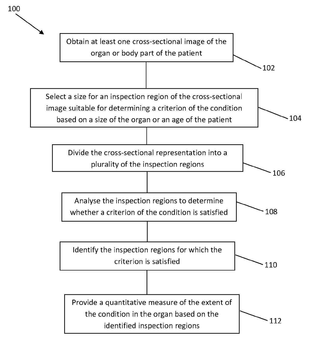
[0055]FIG. 1 illustrates a method 100 of assessing the extent of a condition in an organ or body part of a patient. In a specific embodiment, which will be described herein, the condition is cystic fibrosis and the organ is a lung. However, the method 100 is not limited to this embodiment, and may be used for assessing other conditions, such as disorders causing immune deficiency, primary ciliary dyskinesia, and non-cystic fibrosis bronchiectasis, or diseases in other organs and body parts.

[0056]The method 100 may be used as a measuring tool for assisting in the diagnosis, monitoring the progression of cystic fibrosis, or to evaluate the effectiveness of drugs in clinical trials. For example, the method may be repeated after a period of time and at discrete time intervals such that a progression or treatment of the disease can be monitored. Thus, the method may be used to identity or establish a marker or indicator associated with the condition to assist in determining whether or no...


PUM


Abstract

A method is disclosed for analysing an image to assess a condition of an organ of a patient represented in the image. The method includes initially selecting a spatial resolution for an inspection matrix having a number of inspection regions each delimiting a part of the image. The inspection matrix is applied to the image and the part of the image within each of the inspection regions is analysed to determine a condition thereof. The condition is compared with a predetermined criterion and, if the condition is deemed to satisfy the criterion, the inspection region is identified, e.g. by annotating it with a colour. Finally a quantitative measure of an extent of the condition in the image is provided that is based on a number of the identified or annotated inspection regions.

Description

FIELD OF THE INVENTION[0001]The present invention relates to a method of analysing an image for assessing a condition of an organ of a patient. By way of example only, the method may be used to assess the extent of symptoms of cystic fibrosis in a lung of a patient.BACKGROUND[0002]Standardised outcome measures are in one sense a measuring tool for diagnosis, monitoring disease progress, or to evaluate the effectiveness of certain treatments in medical research. For example, standardised outcome measures are used in clinical trials to test the effectiveness of a certain drug being trialled on a patient over a period of time. It is important that standardised outcome measures are repeatable and can produce reliable results.[0003]Currently there are no standardised outcome measures appropriate for very young children with cystic fibrosis. This effectively excludes infants and young children from clinical trials in an era where new, potentially disease modifying drugs, are becoming avai...

Claims


Application Information

Patent Timeline
Patent Type & Authority Applications(United States)
IPC IPC(8): G06T7/00G06T7/11
CPCG06T7/0016G06T7/11G06T2207/10081G06T2207/10088G06T2207/10012G06T2207/30061A61B6/5217A61B5/055A61B5/7267G06T7/0012G06T2207/20021
Inventor STICK, STEPHENROSENOW, TIMTIDDENS, HARMANNUS ARNOLDUS WILHELMUS MARIADE BRUIJNE, MARLEEN
Owner ERASMUS UNIV MEDICAL CENT ROTTERDAM ERASMUS MC
Why Eureka
Try Eureka