
Shock wave balloon catheter with insertable electrodes

a balloon catheter and electrode technology, applied in balloon catheters, medical science, surgery, etc., can solve the problem of not having a sufficient diameter to accommodate a lithoplasty® apparatus, and achieve the effect of complete dilation of the blood vessel and softening the calcified lesions

Active Publication Date: 2018-06-07
SHOCKWAVE MEDICAL
View PDF5 Cites 22 Cited by
  • Summary
  • Abstract
  • Description
  • Claims
  • Application Information

AI Technical Summary

Benefits of technology

The patent describes a new treatment for small blood vessels with calcified lesions that can be caused by atherosclerosis. Many existing therapies are appropriate for larger vessels, but smaller ones may not have a sufficient diameter to accommodate conventional devices. This patent proposes a treatment using a collapsed angioplasty balloon that is designed to fit into narrow blood vessels and can be inflated with a conducting fluid to pre-dilate the vessel before applying shock wave therapy. The treatment involves the use of a translatable electrode carrier that delivers shock waves to the hardened plaques in the blood vessel walls to soften them. This allows for easier dilation of the vessel. In heavily occluded blood vessels, the treatment may be repeated multiple times to first pre-dilate the vessel and then use the angioplasty balloon for further dilation. The patent is important because it offers a new and effective treatment for small blood vessels that may have been previously treated with traditional therapies.

Problems solved by technology

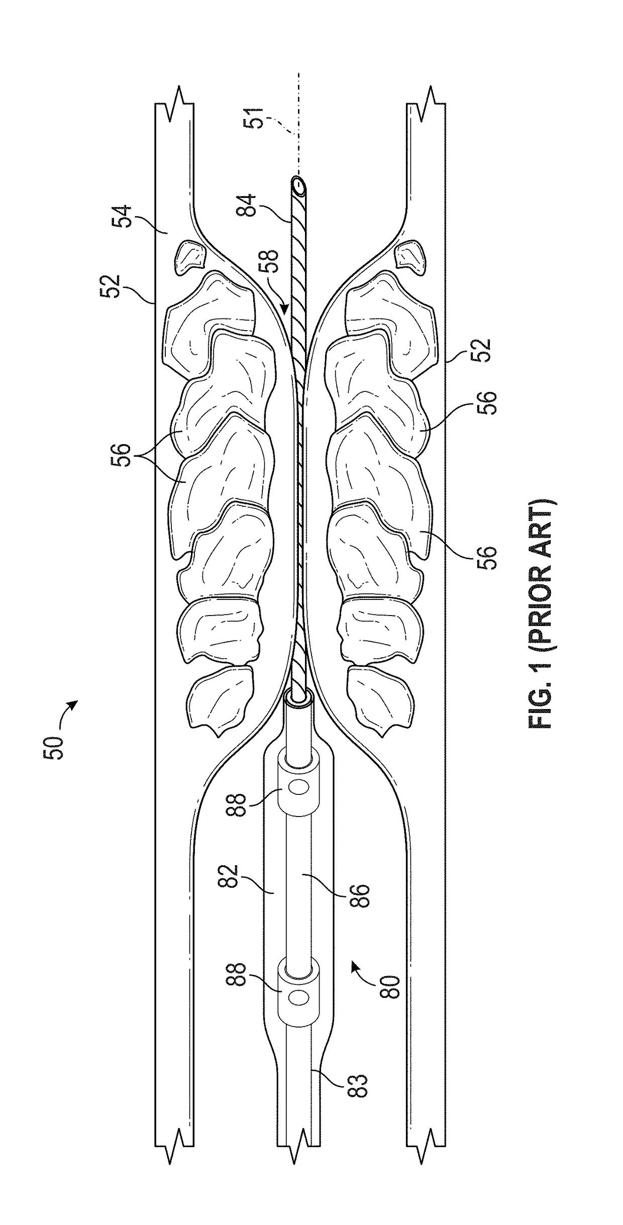
While existing cardiovascular intervention therapies are appropriate for treating calcified lesions in larger blood vessels, e.g., leg arteries having diameters of about 5.0-10.0 mm, smaller vascular structures, or those that are severely blocked may not have a sufficient diameter to accommodate a Lithoplasty® apparatus.

Method used


Image

  • Shock wave balloon catheter with insertable electrodes
  • Shock wave balloon catheter with insertable electrodes
  • Shock wave balloon catheter with insertable electrodes

Examples


first embodiment

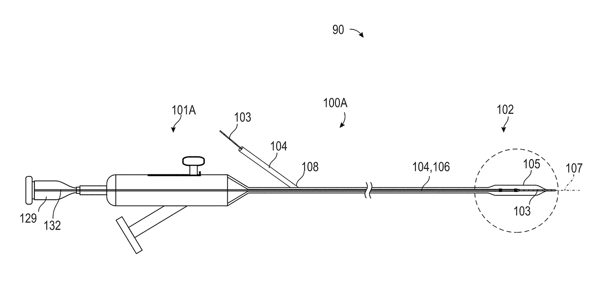
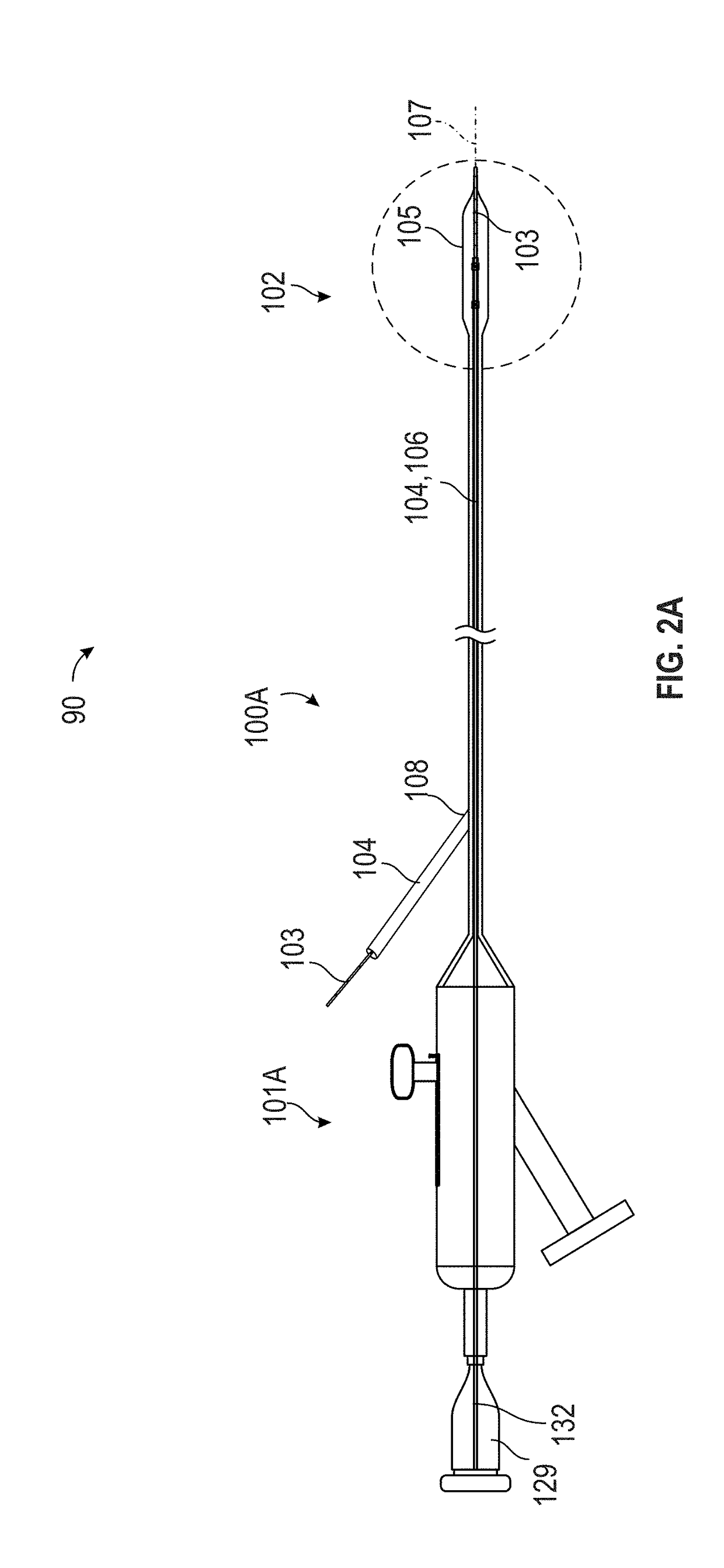
[0030]FIG. 2A illustrates the translatable shock wave treatment apparatus 90, in which the guide wire 103 joins the catheter system 100A at a sealed entry port 108. The catheter system 100A is known in the art as a rapid exchange (Rx) system. The handle assembly 101A includes an electrical connector 129 that transmits electrical power to the emitter assembly 102 via wires 132. The handle assembly 101A is configured to translate the emitter assembly 102 with respect to the angioplasty balloon 105.

second embodiment

[0031]FIG. 2B illustrates the translatable shock wave treatment apparatus 90, in which the guide wire 103 extends all the way through the catheter system 100B, including the handle assembly 101B. The catheter system 100B is known in the art as an over-the-wire system. The handle assembly 101B includes a guide wire handle 128 that can be used to extend and retract the guide wire 103. The handle assembly 101B also includes an electrical connector 129 that transmits electrical power to the emitter assembly 102 via the wires 132. Either one of the rapid exchange system 100A or the over-the-wire system 100B can be equipped with the emitter assembly 102, the electrode carrier 106, and either handle assembly 101A, or 101B to permit translation of the emitter assembly 102 into and out of the angioplasty balloon 105.

[0032]FIGS. 3A and 3B show magnified views of the distal end of the translatable shock wave treatment apparatus 90, in which the guide wire lumen 104 supports the emitter assembl...


PUM


Abstract

A translatable shock wave treatment apparatus is suitable for use in treating calcified lesions in vascular structures having small diameters. An elongate member carrying a collapsed angioplasty balloon is first inserted into the occluded blood vessel. The angioplasty balloon is inflated with a conducting fluid to pre-dilate the narrow blood vessel prior to introducing electrodes and applying shock wave therapy. After the blood vessel is at least partially opened, a translatable electrode carrier equipped with one or more shock wave emitters is advanced into the angioplasty balloon. Shock waves are then propagated through the fluid to impart energy to calcified plaques along the vessel walls, thereby softening the calcified lesions. Following the shock wave treatment, multiple inflation and deflation cycles of the angioplasty balloon can be administered to gently compress the softened lesion and complete dilation of the blood vessel.

Description

FIELD[0001]The present disclosure relates generally to occlusions in human vascular structures and, in particular, to the use of shock wave therapy in the treatment of calcified lesions.BACKGROUND[0002]Balloon angioplasty is a well-known, standard treatment that restores blood flow in blocked arteries. Blockages occur when plaque accumulates in the walls of the blood vessels, forming lesions. In a conventional balloon angioplasty procedure, a catheter carrying an angioplasty balloon is inserted into the blood vessel along a guide wire to position the angioplasty balloon adjacent to a lesion. Progress of the guide wire may be tracked using fluoroscopy or x-rays. Inflating the angioplasty balloon compresses soft lesions on the wall of the vessel, thereby dilating the blood vessel and allowing blood to flow through a larger portion thereof. However, when plaque is hardened, or calcified, gentle compression may not be effective. In such cases, rapid expansion of the angioplasty balloon ...

Claims


Application Information

Patent Timeline
Patent Type & Authority Applications(United States)
IPC IPC(8): A61B17/22A61M25/10
CPCA61B17/22022A61M25/104A61M2025/0008A61B2017/22025A61B2017/22051A61B17/22012
Inventor KAT-KUOY, DENNY
Owner SHOCKWAVE MEDICAL
Learn More