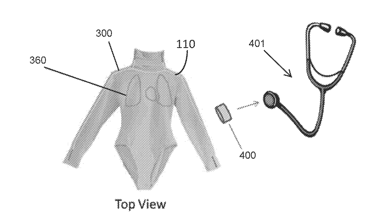
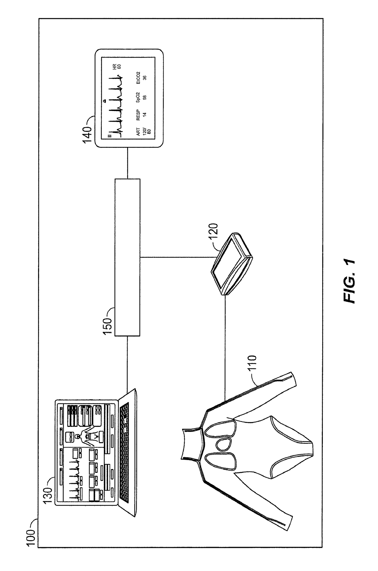
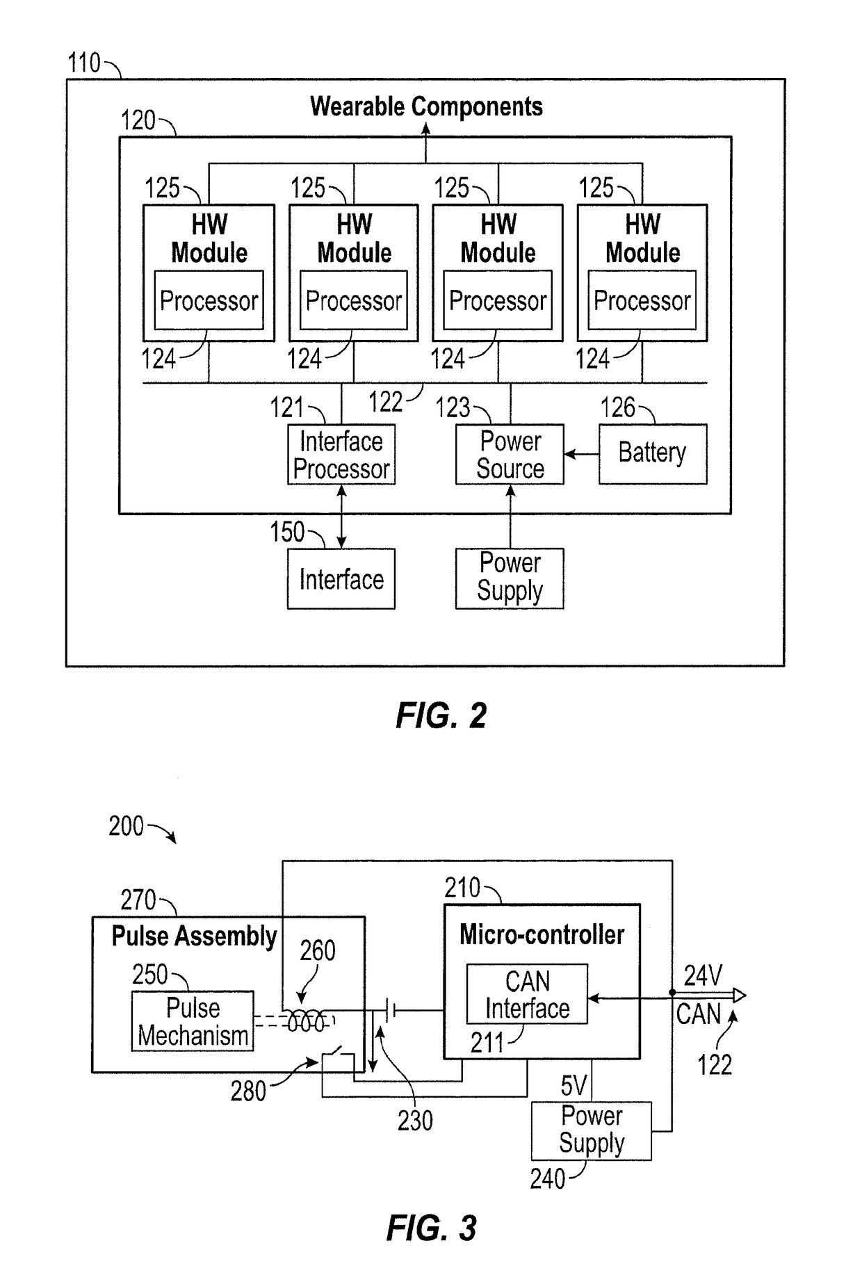
Unfortunately, deliberate practice of medical procedures is not easy to obtain due to the intrinsic hazards and complexities of
patient care.
Many of the existing publications related to wearable
simulation garments describe possible patient parameters that could be simulated but fail to provide any meaningful teaching as to how to accomplish related functionality.
Existing wearable simulation garments are inadequate for use in a training environment for at least the following reasons:1. the parameters provided are not good representations of actual
physiology;2. the real parameters from the standardized patient are not isolated from the simulated parameters compromising the simulation;3. the simulated parameters are discomforting to the standardized patient (e.g., motion,
noise, weight);4. the components required to be located on the patient are large and / or bulky making them difficult to wear and conceal;5.
power consumption of the system is too high to allow for a body worn battery and could generate uncomfortable heat; and6. connections are required to non-body worn components that could
restrict the movement of the standardized patient and could also result in pinched tubes making the solution inoperable.
It does not appear that a wearable simulation garment has been accepted in the marketplace (or perhaps never even made it to market) likely due to these problems.
While this approach allows for a self-contained, highly mobile solution, it tends to result in an expensive solution that is not easily scalable and may be difficult to operate and maintain.
Organizations providing
medical training (e.g., hospitals, medical schools, and
nursing schools) frequently have a mix of these manikin-based simulator products and availability of standardized patients to satisfy
training needs, which results in significant cost,
physical space requirements, and maintenance difficulties.
Additionally, there is a large gap in the mid-range of the price and performance curve for medical simulators.
Users are often forced to choose between low to mid-priced products focused on procedural training or high-end expensive products focused on
cognitive training.
There is little compatibility and
interoperability between products, and there is limited
modularity and configurability of individual products.
For example, physical
modularity in current products is typically limited to optional limbs (e.g., IV arm,
blood pressure arm, trauma limbs, etc.) or interchangeable genitalia.
As a result, simulators cannot be interchanged or easily configured for different
training needs.
Another common issue with standardized patient simulation and manikin-based medical simulators is the generation of body sounds, such as heart, lungs, and bowel sounds.
However, none of these techniques produce a reliable, cost-effective and automatic means of creating realistic body sounds and are not suitable for use in wearable components.
However, this approach has many drawbacks and limitations.
For example,
sound quality can be poor due to resonances and vibrations in the manikin, and the low-end
frequency response can be poor due to limited speaker size.
In addition, localizing sounds to a particular area of the manikin can be difficult since sounds travel within the manikin.
Further,
noise from other system components, such as motors and solenoids, can easily be picked up with the
stethoscope.
In addition, this technique does not transfer well for implementation in a wearable solution for a standardized patient.
However, speakers of adequate bandwidth to reproduce body sounds tend to be large and bulky making them difficult to conceal and wear.
The
sound field is difficult to control and it is likely that actual body sounds from the standardized patient would be heard unless there was adequate sound damping material that would make the garment bulky and unrealistic.
In addition, sound insulating materials are typically thermal insulators and would make the garment hot and uncomfortable for the standardized patient.
In addition, a special
stethoscope must be used that is capable of receiving the transmitted sound or control signals which further increases the cost and complexity of the system.
Additionally, this technique requires constant attention from an instructor and prohibits standalone use by a trainee.
However, the realism, limitations, and cost of these simulated physiological functions varies greatly depending on the particular implementation.
However, none of these techniques produce a reliable, cost-effective, and realistic pulse and are not suitable for use in wearable components for a standardized patient.
However, the compressor or pump adds significant cost, increases
power consumption, and can create undesirable
noise.
In addition, valves need to be used if the different pulse points need to be controlled separately, thereby adding to the cost and complexity of the implementation.
There is no place to locate these components in a wearable simulation garment that would be unobtrusive.
The components could be located in a separate
enclosure, but that implementation would require tubes running from the
enclosure to the garment, and could
restrict the movement of the standardized patient or result in pinched tubes.
However, the resulting pulse tends to feel artificial due to the rigidity of the simulated
artery and / or the vertical movement, rather than a flowing and expanding movement.
A compressor is bulky, adds significant cost, increases
power consumption, and can create noise.
Emptying the bladder is typically done with a bleeder valve which adds more cost.
There is no place to locate these components in a wearable simulation garment that would be unobtrusive.
Connected tubes could
restrict the movement of the standardized patient and could result in pinched tubes making the solution inoperable.
Additionally, isolating the
breathing motion of the standardized patient is not possible with current solutions.
Accordingly, none of the above described techniques produce a reliable, cost-effective and automatic means of creating realistic simulation parameters (e.g., body sounds, pulses, and
breathing motions) to add high-fidelity features to lower fidelity manikin simulators and to standardized patients.


