
Non-human animal having human cd3 gene substituted for endogenous cd3 gene

a technology of human cd3 and endogenous cd3 gene, which is applied in the field of human cd3 gene substitution of nonhuman animals, can solve the problems of inability to show hla-restricted immune response, human hematocyte differentiation is not completely reproduced, and the therapeutic antibodies having an additional function to activate t cells are difficult to evaluate through animal tests in preclinical studies

Active Publication Date: 2018-07-12
CHUGAI PHARMA CO LTD
View PDF2 Cites 6 Cited by
  • Summary
  • Abstract
  • Description
  • Claims
  • Application Information

AI Technical Summary

Benefits of technology

The patent text describes a genetically modified non-human animal that is deficient in certain genes and expresses human genes to functionally replace them. This results in an animal that can be used to evaluate human CD3-based therapeutic agents and methods. The technical effect of this invention is the ability to develop highly specific human CD3-based therapies using genetically modified non-human animals.

Problems solved by technology

However, therapeutic antibodies having an additional function to activate T cells as mentioned above are difficult to evaluate through animal tests in preclinical studies.
However, in such model animals, since the cytokines, growth factors and microenvironment-forming cells that are necessary for proliferation and differentiation of human hematopoietic stem cells all remain mouse-derived, human hematocyte differentiation is not reproduced completely.
Recently, further improvements have been achieved by humanization of interleukin-3 (IL-3), GM-CSF, and Stem Cell Factor (SCF), but there still remains the problem that T cells are unable to show HLA-restricted immune response (Non-patent Document 7).
However, such transgenic mice show marked decrease in the number of thymocytes, and it is difficult to say that they are mice with normal immune function.
Furthermore, there is no report on genetically modified mice in which the full lengths of CD3ε, CD3δ, and CD3γ, spanning the extracellular region through the cytoplasmic region, have been humanized.

Method used


Image

  • Non-human animal having human cd3 gene substituted for endogenous cd3 gene
  • Non-human animal having human cd3 gene substituted for endogenous cd3 gene
  • Non-human animal having human cd3 gene substituted for endogenous cd3 gene

Examples


example 1

n of Human CD3 Gene-Substituted Mice

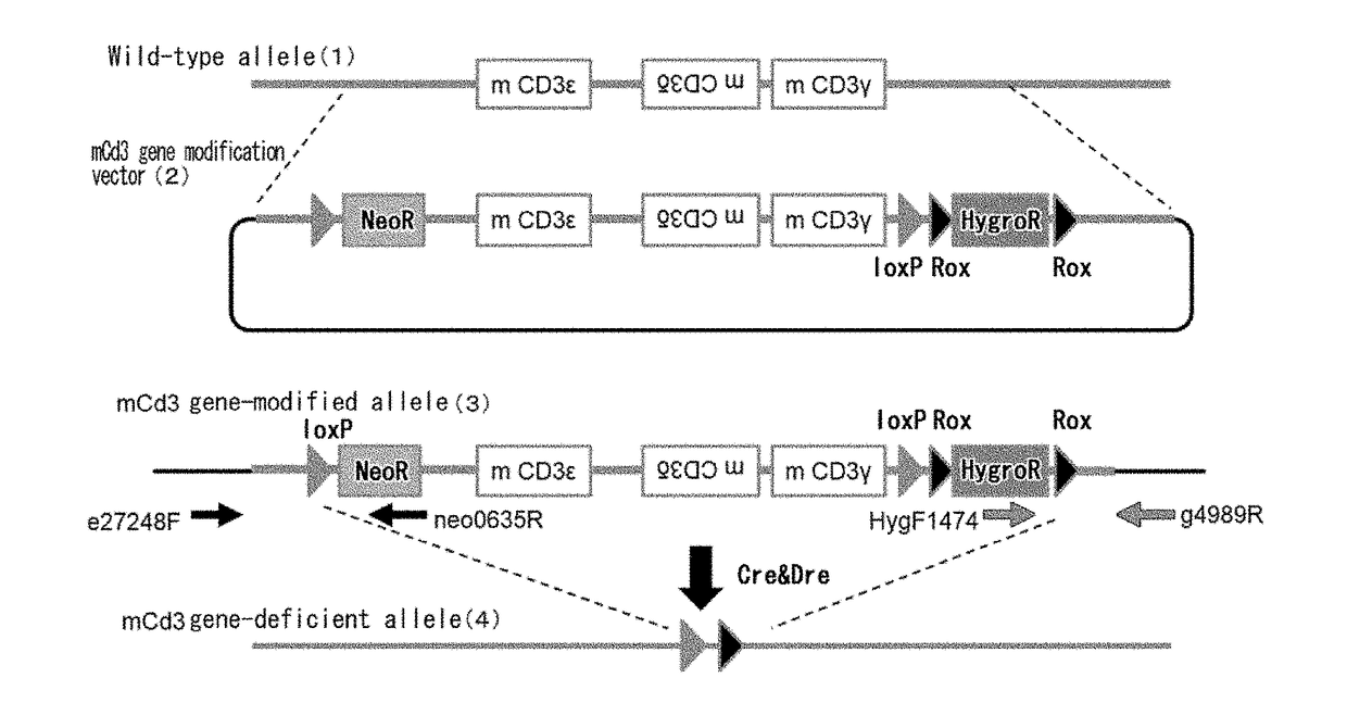
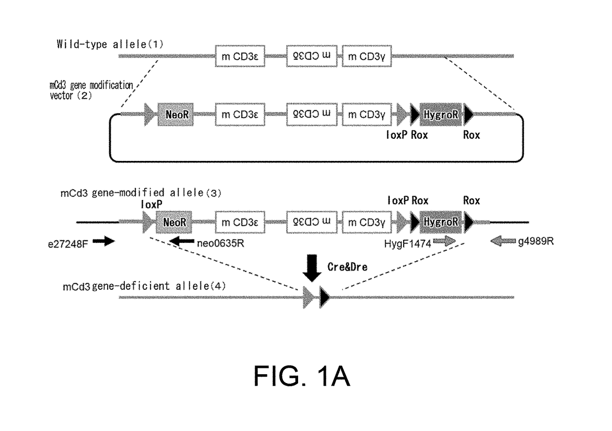
[0305](1) Construction of a Mouse Cd3 Gene Region Modification Vector (FIG. 1A)

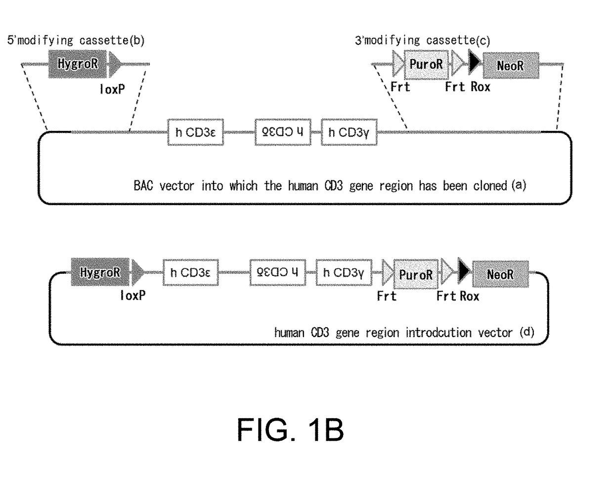
[0306]A bacterial artificial chromosome (BAC) clone was used, into which a genomic region where the mouse CD3ε, CD3δ, and CD3γ genes are positioned had been cloned. A loxP sequence was inserted at the position approximately 3.5 kb 5′ upstream of the gene region encoding mouse Cd3ε in this BAC, and the genome region further upstream was removed leaving approximately 3.1 kb. At that time, the loxP sequence was introduced together with neomycin-resistance (neo) gene cassette and insertion was conducted by homologous recombination using a Red / ET system (GeneBridges). In that case, from among the Escherichia coli clones that grew in a kanamycin-supplemented medium, clones for which polymerase chain reaction (PCR) method resulted in correct amplification were selected. Next, loxP sequence and Rox sequences were placed at 3′ downstream of the Cd3γ gene on the BAC. More specifi...

example 2

n of Cell Proliferation Ability of Spleen Cells in Human Cd3 Gene-Substituted Mice

[0333]Spleens were collected from mice (12-week old, male), and cells were isolated using 70 μm mesh. Erythrocytes were lysed by adding a hemolytic agent (manufactured by SIGMA). Phytohaemagglutinin (PHA, manufactured by SIGMA) which is a T cell mitogen was added to the isolated spleen cells, and after culturing for five days, MTS assay was carried out using a cell proliferation assay reagent (Cell Titer 96. Aqueous One Solution Reagent, manufactured by Promega). Cell proliferation activities of the respective genotypes are shown as relative proportions by defining the activity without mitogen addition as 1 (FIG. 9).

[0334]As a result, in spleen cells of the Cd3 gene-deficient mice, cell proliferation activity in response to mitogen stimulation tended to decrease compared to that when mitogen was not added, and was approximately 60% of that of wild-type mice. On the other hand, in spleen cells of the hu...

example 3

n of Cytokine Production in Spleen Cells of Human CD3 Gene-Substituted Mice

[0335]Spleen cells were prepared by collecting spleens from mice, isolating cells using 70 μm mesh, and lysing erythrocytes by adding a hemolytic agent. After culturing the cells for three days in the medium supplemented with phytohaemagglutinin (PHA) which is a T cell mitogen at a final concentration of 1 μg / mL, cytokines produced in the medium were measured (FIG. 10).

[0336]As a result, in spleen cells of the Cd3 gene-deficient mice, mitogen stimulated-cytokine production was not observed, whereas in the human CD3 gene-substituted mice, cytokine production was observed, indicating that the function of producing cytokines was restored.


PUM

PropertyMeasurementUnit
concentrationaaaaaaaaaa

Abstract

The present invention provides genetically modified non-human animals which are deficient in at least one or more types of CD3 genes selected from the group consisting of endogenous CD3ε, CD3δ, and CD3γ in its genome and functionally express at least one or more types of human CD3 genes selected from the group consisting of human CD3ϵ, CD3δ, and CD3γ. In the genetically modified non-human animals of the present invention, mature T cell differentiation and production can take place, and immunocompetent cells including T cells can exert their functions. The genetically modified non-human animals of the present invention enable efficient evaluation and screening in the development of therapeutic agents and therapeutic methods that use human CD3-mediated targeted drugs.

Description

TECHNICAL FIELD[0001]The present invention relates to human CD3 gene-substituted non-human animals, and methods for evaluating compounds using the human CD3 gene-substituted non-human animals, and such.BACKGROUND ART[0002]A T-cell receptor (TCR) complex is an antigen-receptor molecule expressed mainly on T cells. It is composed of antigen-binding TCRα and β chains (or δ and γ chains), and multiple CD3 molecules (CD3-epsilon (ε), CD3-delta (δ), and CD3-gamma (γ)) and CD247 that mainly have a function of transducing signals into cells.[0003]TCRα and TCRβ are formed from variable and constant regions. Through V(D)J gene rearrangement similarly to antibody molecules, the variable regions constitute a repertoire that presents binding activities to various antigen molecules that differ for every cell clone. T cells are activated through specific binding via TCR complexes to antigen molecules presented by major histocompatibility complexes (MHCs) on the cell surface of dendritic cells and ...

Claims


Application Information

Patent Timeline
Patent Type & Authority Applications(United States)
IPC IPC(8): A01K67/027C07K14/725C12N15/85G01N33/15
CPCA01K67/0278C07K14/7051C12N15/8509G01N33/15C12N2015/8518C12N15/09G01N33/5008C07K16/2809A01K2207/12A01K2217/072A01K2227/105A01K2267/0325A01K2267/0331
Inventor JISHAGE, KOICHIUEDA, OTOYAWADA, NAOKOISHIGURO, TAKAHIROKINOSHITA, YASUKO
Owner CHUGAI PHARMA CO LTD
Learn More