Devices and methods for optical pathology

a technology of optical pathology and optical x-ray, applied in the field of optical x-ray devices and methods, can solve the problems of toxic hematoxylin and cannot be applied in vivo, and achieve the effect of improving the discrimination of cancerous tissu

Active Publication Date: 2018-07-26
UNIV OF MASSACHUSETTS
View PDF1 Cites 0 Cited by
  • Summary
  • Abstract
  • Description
  • Claims
  • Application Information

AI Technical Summary

Benefits of technology

[0015]Reflectance and fluorescence images can be processed to form combined or pseudo color images to provide enhanced discrimination of cancerous tissue. The system has a resolution sufficient to provide for the detection of abnormal tissue at the cellular level. Thus individual cells within a subregion of tissue can be quantatively characterized as normal or abnormal using the fluorescence polarization methods described herein. Fluorescence polarization images of tumor cells were obtained and quantitative values computed on a numeric scale that distinguished between cancerous and normal tissue. Autofluorescence images can also be obtained to further assist in diagnosis of certain conditions.

Problems solved by technology

Hematoxylin is toxic and cannot be applied in vivo, whereas eosin is approved for human use.

Method used


Image

  • Devices and methods for optical pathology
  • Devices and methods for optical pathology
  • Devices and methods for optical pathology

Examples


Embodiment Construction

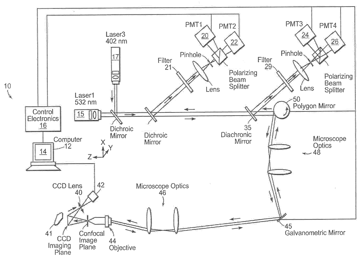
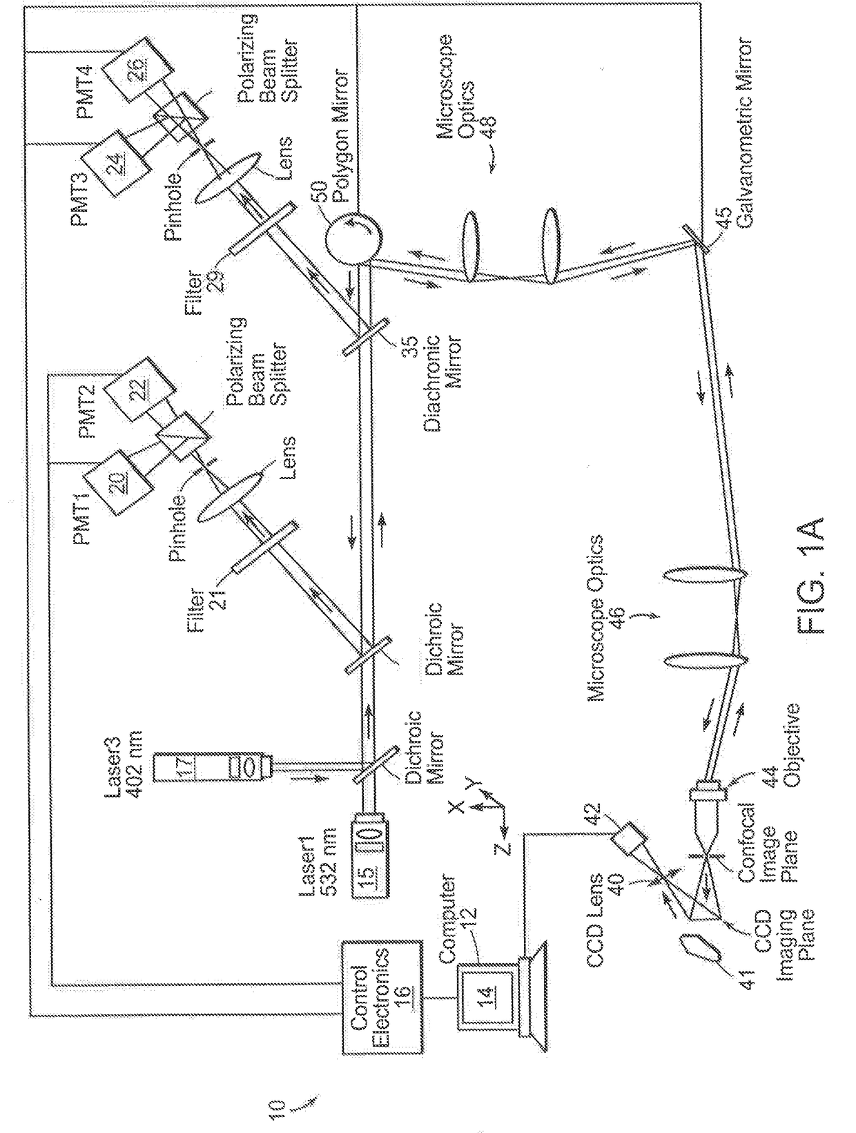
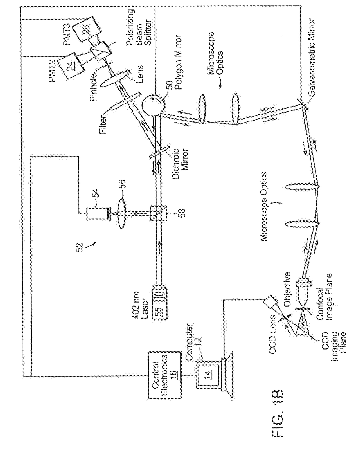
[0037]A method and device for intraoperative, in vivo, detection of pathological cells and margins. Preferred embodiments utilize a plurality of complementing stains that are imaged for delineating cancers such as skin or breast cancers during surgery.

[0038]Clinical evidence shows that eligible patients undergoing breast conservation therapy (BCT) have the same long-term survival rate as those undergoing mastectomy, when patients with BCT do not have a local regional recurrence. Thus, complete removal of breast cancer is of primary importance. Currently, re-excision is required in up to 60% cases because most of BCT procedures are performed without intraoperative margin control. Standard histopathological evaluation of the excision margins performed during the surgery reduces re-excision rate to approximately 20%. However, due to high cost, it has not become widely available. Therefore, a reliable cost-effective method for real-time examination of breast cancer margins and other can...


PUM


Abstract

Currently most cancers, including breast cancers, are removed without any intraoperative margin control. Post-operative methods inspect 1-2% of the surgical margin and are prone to sampling errors. The present invention relates to an optical imaging system that will enable evaluation of the surgical margin in vivo and in real-time. The invention provides for simultaneous fluorescence and fluorescence polarization imaging. The contrast of the acquired images will be enhanced using fluorescent agents approved for diagnostic use in patients. As the staining pattern of fluorescence images is similar to that of histology, and the values of fluorescence polarization are significantly higher in cancerous as compared to normal cells, the invention provides for further improvements in diagnostic methods. The systems and methods can be applied to the intra-operative delineation of cancerous tissue.

Description

CROSS REFERENCE TO RELATED APPLICATIONS[0001]This application is a divisional of U.S. application Ser. No. 14 / 000,106, filed Aug. 16, 2013, which is a U.S. Application National Stage filed under 31 USC 371 of International Application No. PCT / US2012 / 025678, filed Feb. 17, 2012, which claims the benefit of U.S. Provisional Application No. 61 / 443,931, filed Feb. 17, 2011, the entire contents of the above application is incorporated herein by reference.BACKGROUND OF THE INVENTION[0002]There currently exists no intraoperative in situ methods for detecting and delineating pathology. Currently, in the majority of cases, except for the visual assessment of where to excise, cancers are removed without intraoperative margin control. After the surgery is completed and the resulting wound is closed, the tissue is sent for histopathological analysis. Postoperative methods of cancer delineation involve sampling and examine only 1-2% of the surgical margin. For example, the “bread loaf” method us...

Claims


Application Information

Patent Timeline
Patent Type & Authority Applications(United States)
IPC IPC(8): A61B5/00G06T7/90A61M31/00A61M5/00G06T7/00
CPCG06T7/90G06T2207/30096G06T2207/20221G06T2207/10152G06T2207/10064A61B5/0071A61B5/0091A61M31/005A61M5/007A61B5/742A61B5/0068G06T7/0012
Inventor YAROSLAVSKY, ANNA N.PATEL, RAKESHWIRTH, DENNIS
Owner UNIV OF MASSACHUSETTS
Why Eureka
Try Eureka