Ultrasound and photoacoustic systems and methods for fetal brain assessment during delivery

a technology applied in the field of ultrasound and photoacoustic system and methods for fetal brain assessment during labor and delivery, can solve the problems of reduced oxygen availability to body tissues, brain damage to the fetus, and deprived fetus of oxygen supply

Active Publication Date: 2018-08-02
WAYNE STATE UNIV
View PDF1 Cites 9 Cited by
  • Summary
  • Abstract
  • Description
  • Claims
  • Application Information

AI Technical Summary

Benefits of technology

[0008]According to the subject matter of the present disclosure, a system for fetal brain assessment during delivery may include one or more processors, one or more memory modules communicatively coupled to the one or more processors, an ultrasound machine comprising a display and communicatively coupled to the one or more memory modules, a probe device communicatively coupled to the ultrasound machine, and machine readable instructions stored in the one or more memory modules. The machine readable instructions may cause the system to perform at least the following when executed by the one or more processors: transmit a plurality of (ultrasound) US and (photoacoustic) PA signals (i.e., safe

Problems solved by technology

Intrapartum hypoxia, or fetal hypoxia, may occur during delivery of the fetus, causing the fetus to be deprived of an adequate supply of oxygen.
Intrapartum hypoxia results in a diminished availability of oxygen to body tissues despite adequate perfusion of the tissue by blood.
Such a condition may also result in brain damage to the fetus, including short and/or long term neurological complications.
Further, such a condition may be life threatening unless prompt actions are taken to restore well-oxygenated blood to the fetus.
Many cesarean sections are performed, however, without clear documentation of the fetal status.
However, a fetus may have a placental reservoir t

Method used


Image

  • Ultrasound and photoacoustic systems and methods for fetal brain assessment during delivery
  • Ultrasound and photoacoustic systems and methods for fetal brain assessment during delivery
  • Ultrasound and photoacoustic systems and methods for fetal brain assessment during delivery

Examples


Embodiment Construction

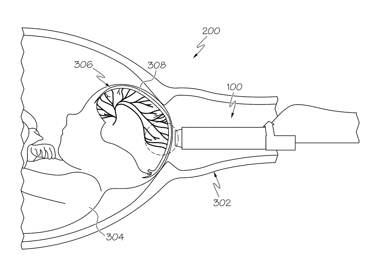
[0022]The present disclosure relates to systems and methods to optimize clinical care of a fetus and mother care in labor during active labor and delivery by providing direct information regarding oxygen saturation in arterial and venous fetal cerebral and / or cortical blood to monitor the fetal status for intrapartum hypoxia. The systems and methods described herein further permit a visualization of fetal brain tissue and vessels to estimate blood flow and global oxygen consumption in the visualized fetal brain vessels. Further, the systems and methods described herein provide for an estimation of blood movements as a proxy of regional blood perfusion and direction an ultrasound (US) visualization of a pose (i.e., position and orientation) of a head of the fetus in the maternal pelvis during active labor and delivery.

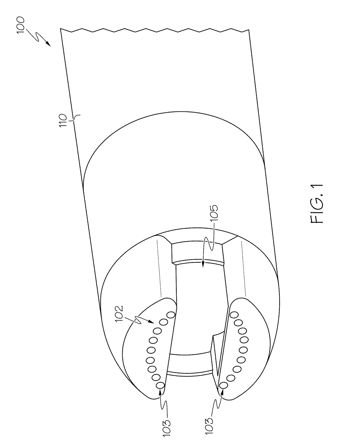
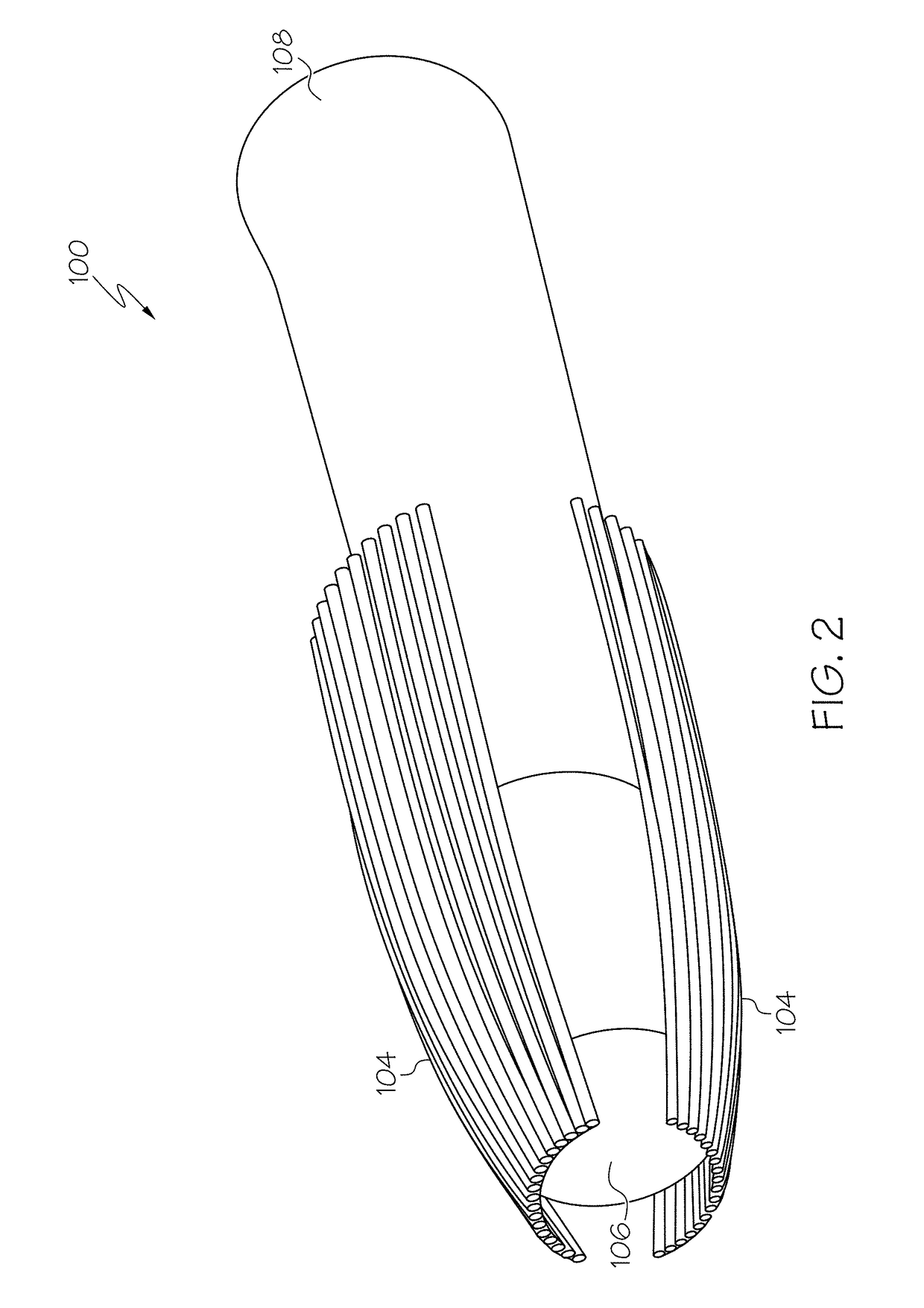
[0023]Referring initially to FIGS. 1-2, a probe device 100 illustrated for assessment of the an oxygen measurement in the fetal brain during labor and delivery of a fet...


PUM


Abstract

Methods and system are described for multi-parametric, non-invasive, and real-time assessment of blood perfusion and oxygenation in the fetal brain during labor and delivery of a fetus through a vaginal birth canal of a maternal pelvis, and include positioning a probe device in the maternal pelvis during active labor, transmitting and receiving a plurality of ultrasound (US) and photoacoustic (PA) signals between the probe device and fetal brain, displaying in real-time on an US machine communicatively coupled to the probe device one or more images of venous and arterial blood flow of respective blood vessels in the fetal brain, measuring oxygen saturation of the respective venous and arterial blood vessels based on data from the one or more images, and estimating the oxygen measurement in the fetal brain during active labor based on the measured oxygen saturation.

Description

CROSS REFERENCE TO RELATED APPLICATIONS[0001]The present specification claims priority to U.S. Provisional App. No. 62 / 451,392, filed Jan. 27, 2017, and entitled “ULTRASOUND AND PHOTOACOUSTIC SYSTEMS AND METHODS FOR FETAL BRAIN ASSESSMENT DURING DELIVERY,” the entirety of which is incorporated by reference herein.TECHNICAL FIELD[0002]The present disclosure generally relates to ultrasound (US) and photoacoustic (PA) systems and methods for fetal brain assessment during labor and delivery and, more specifically, to US and PA systems and methods for multi-parametric, non-invasive, and real-time assessment of oxygen consumption in the fetal brain during labor and delivery of a fetus through a vaginal birth canal.BACKGROUND[0003]Intrapartum hypoxia, or fetal hypoxia, may occur during delivery of the fetus, causing the fetus to be deprived of an adequate supply of oxygen. Intrapartum hypoxia results in a diminished availability of oxygen to body tissues despite adequate perfusion of the t...

Claims


Application Information

Patent Timeline
IPC IPC(8): A61B8/08A61B5/00A61B8/12A61B5/1455
CPCA61B8/0866A61B8/488A61B8/0891A61B5/0095A61B8/0808A61B8/12A61B5/14552A61B5/1464A61B5/4064A61B5/4362A61B2503/02
Inventor MEHRMOHAMMADI, MOHAMMADHERNANDEZ-ANDRADE, EDGARGELOVANI, JURI G.HASSAN, SONIA S.YAN, YAN
Owner WAYNE STATE UNIV
Why Eureka
Try Eureka