

VE-PTP Extracellular Domain Antibodies Delivered by a Gene Therapy Vector
a technology of extracellular domain and gene therapy, which is applied in the field of ve-ptp extracellular domain antibodies delivered by gene therapy vectors, can solve the problems of visual impairment and blindness, affect the normal structure or function, and affect the normal function
- Summary
- Abstract
- Description
- Claims
- Application Information
AI Technical Summary
Benefits of technology
Problems solved by technology
Method used
Image

Examples
example 1
Identification and Characterization of a VE-PTP Extracellular Domain Binding Agent
[0179]VE-PTP (SEQ ID NO. 15) is the mouse orthologue of HPTPβ. Antibodies to the VE-PTP extracellular domain were identified and characterized as summarized below.
A. Generation of Antibodies to VE-PTP Extracellular Domain Protein (VE-PTP-ECD)
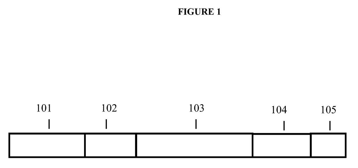
[0180]VE-PTP-Fc fusion protein was constructed such that the first 8 fibronectin type III-like repeats ending with the amino acid proline at position 732 of VE-PTP (SEQ ID NO. 16) were fused in frame with the Fc portion of human IgG1, starting with amino acid proline at position 239. This construct cloned into pcDNA3 was stably transfected into CHO cells, and the fusion protein was purified by protein A Sepharose™ affinity purification.
[0181]The antibody was generated by immunizing rats with the VE-PTP-Fc fusion protein. Immunization, hybridoma-fusion, and screening were conducted using standard methods.
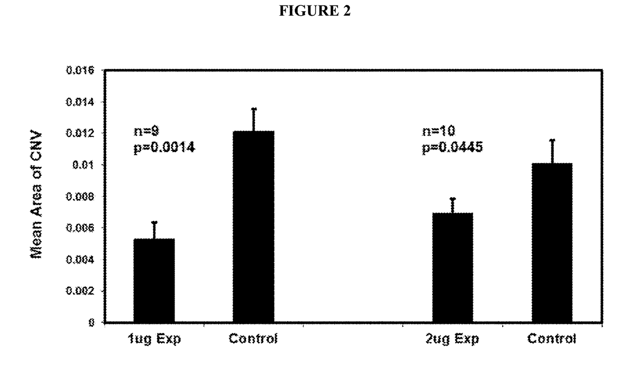
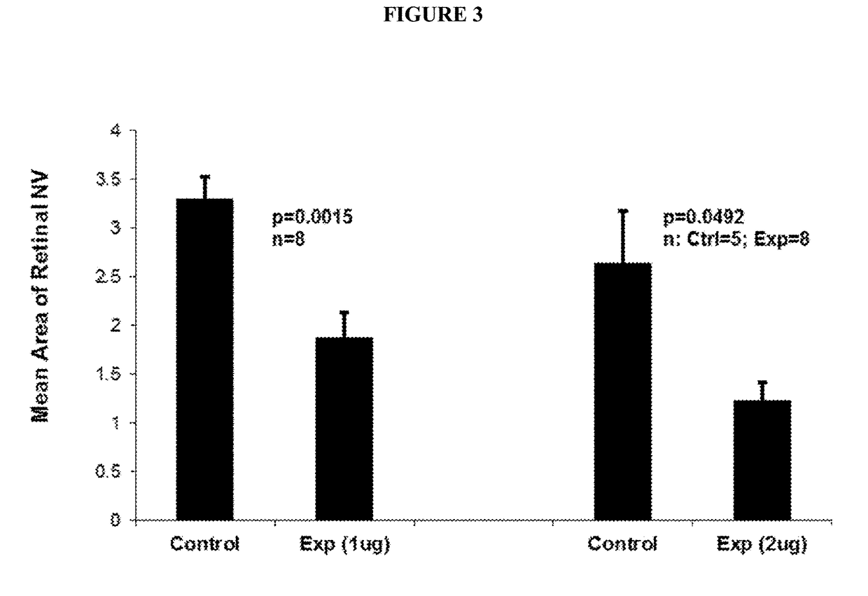
B. Anti-VE-PTP-ECD Activity Studies in Mice Eyes
Laser-Induced ...
example 2
Identification and Characterization of HPTPβ Extracellular Domain Binding Agents
[0184]Antibodies to the HPTPβ extracellular domain (SEQ ID NO. 17) are identified and characterized as summarized below.
Generation of Antibodies to the HPTPβ Extracellular Domain
[0185]An HPTPβ fusion protein is constructed such that the extracellular domain (SEQ ID NO. 17) is fused in frame with the Fc portion of human IgG1, starting with amino acid proline at position 239 (herein referred to as HPTPβ-ECD-Fc). This construct is cloned into pcDNA3 (Invitrogen™ Carlsbad, Calif.) and stably transfected into CHO cells, and the fusion protein is purified by protein A Sepharose™ affinity purification.
[0186]The antibody is generated by immunizing mice with the HPTPβ-ECD-Fc fusion protein. Immunization, hybridoma-fusion, and screening are conducted using standard methods.
Generation of Antibodies to the HPTPβ First FN3 Repeat
[0187]An HPTPβ fusion protein is constructed such that the first FN3 repeat (SEQ ID NO. 1...
example 3
Sequence Analysis of a HPTPβ Extracellular Domain Binding Agent
[0189]The sequence encoding antibody R15E6 was determined; the procedure and results are summarized below.
[0190]Total RNA was extracted from hybridoma cell pellets. Reverse transcription with an oligo(dT) primer was performed to create cDNA from the RNA. The VH and VL regions of R15E6 were amplified from the cDNA using variable domain primers to generate the bands in FIG. 5. The VH and VL products were cloned into the plasmid pCR2.1, transformed into E.coli, and screened by PCR for positive transformants. Positive transformants were analyzed by DNA sequencing. Resulting DNA sequences were compared to determine individual and consensus amino acid sequences of the VH and VL regions. FIGS. 6 and 7 show the individual and consensus amino acid sequence results for the VH and VL regions, respectively.
[0191]From the sequence analysis, consensus amino acid and DNA sequences were determined for the VH (SEQ ID NO.: 1 and SEQ ID NO...
PUM
| Property | Measurement | Unit |
|---|---|---|
| size | aaaaa | aaaaa |
| length of time | aaaaa | aaaaa |
| Thickness | aaaaa | aaaaa |
Abstract
Description
Claims
Application Information