

Anti-cd43 antibody and use thereof for cancer treatment
an anti-cd43 antibody and cancer treatment technology, applied in the field of anti-cd43 antibody and cancer treatment, can solve the problem that anti-cd43 antibodies are not effective in detecting or treating cancer cells
- Summary
- Abstract
- Description
- Claims
- Application Information
AI Technical Summary
Benefits of technology
Problems solved by technology
Method used
Image

Examples
example 1
on of Anti-CD43 Antibody
[0280]1-1. Preparation of Mouse Antibody
[0281]1-1-1. Preparation of Cell Producing Monoclonal Antibody
[0282]It was prepared by the fusion of splenocyte of Balb / c white mouse in which a human thymocyte was injected as an antigen and myeloma cell line SP2 / 0-Ag14 (ATCC, CRL-1581) of 8-azaguanine resistant mouse.
[0283]107 of human thymocytes (Seoul National University Hospital) were intraperitoneally injected into Balb / c white mouse per 2 weeks for 6 weeks to induce an immune response, and the spleen was extracted at 3 days after the last additional inoculation to prepare cell suspension. According to the method of Koeler & Milstein (1975), 108 of splenocytes and 107 of myeloma cells were under cell fusion by using 400 of polyethylene glycol. The fused cells were washed and then suspended in DMEM culture solution supplemented with 100 uM hypoxanthine, 0.44 uM animopterin and 16 uM thymidine (HAT culture solution), and cells were aliquoted in a 96-well plate, and ...
example 2
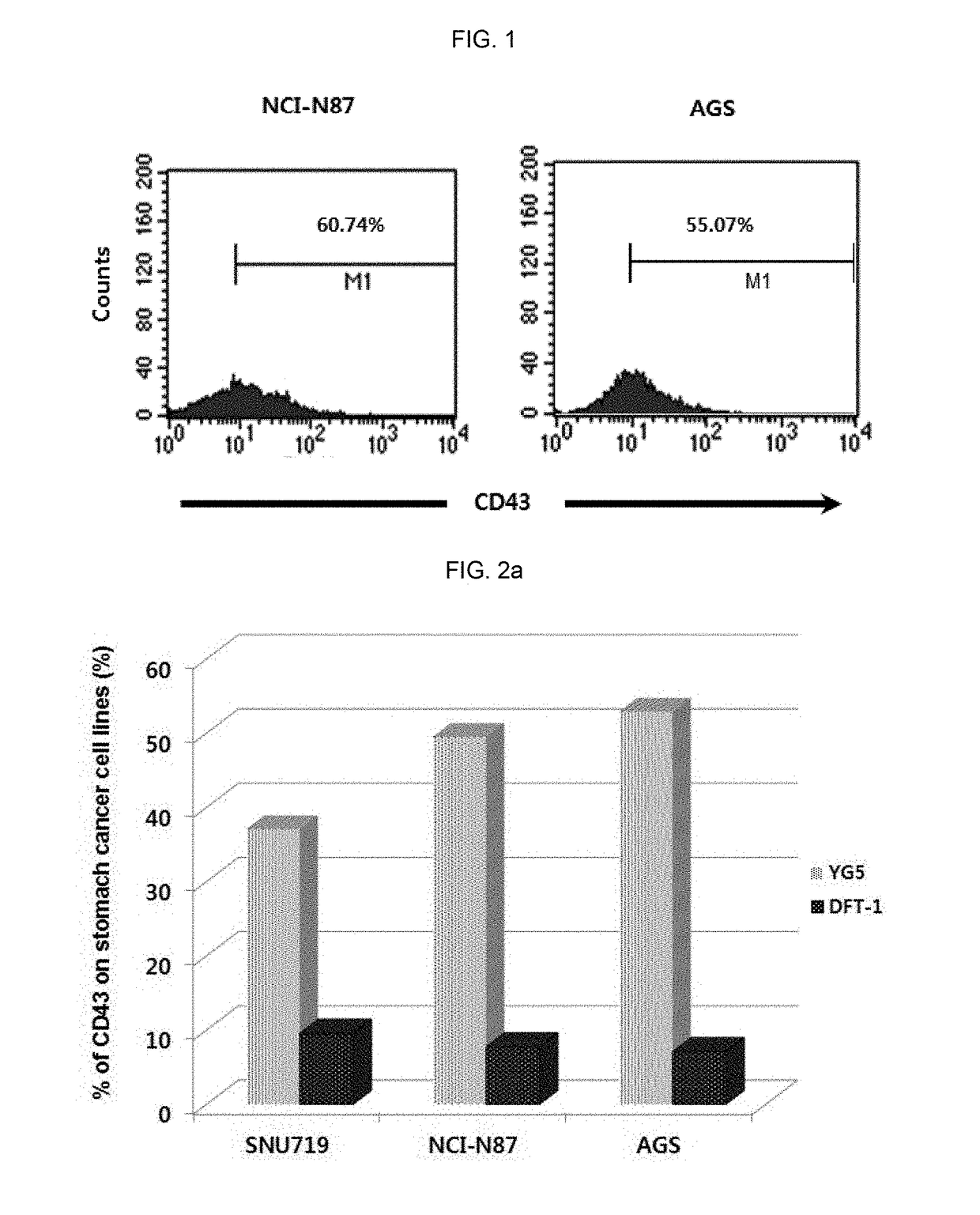
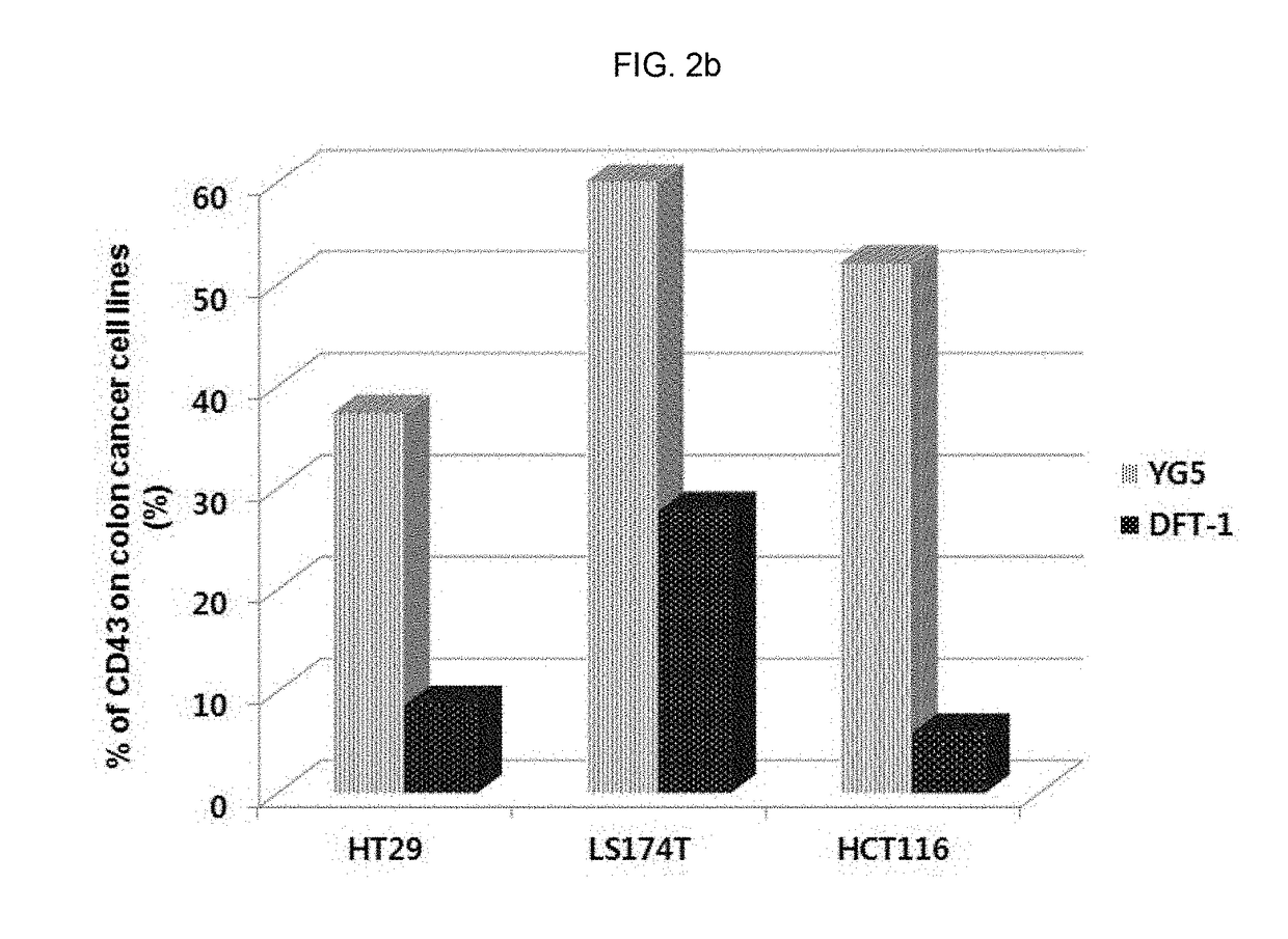
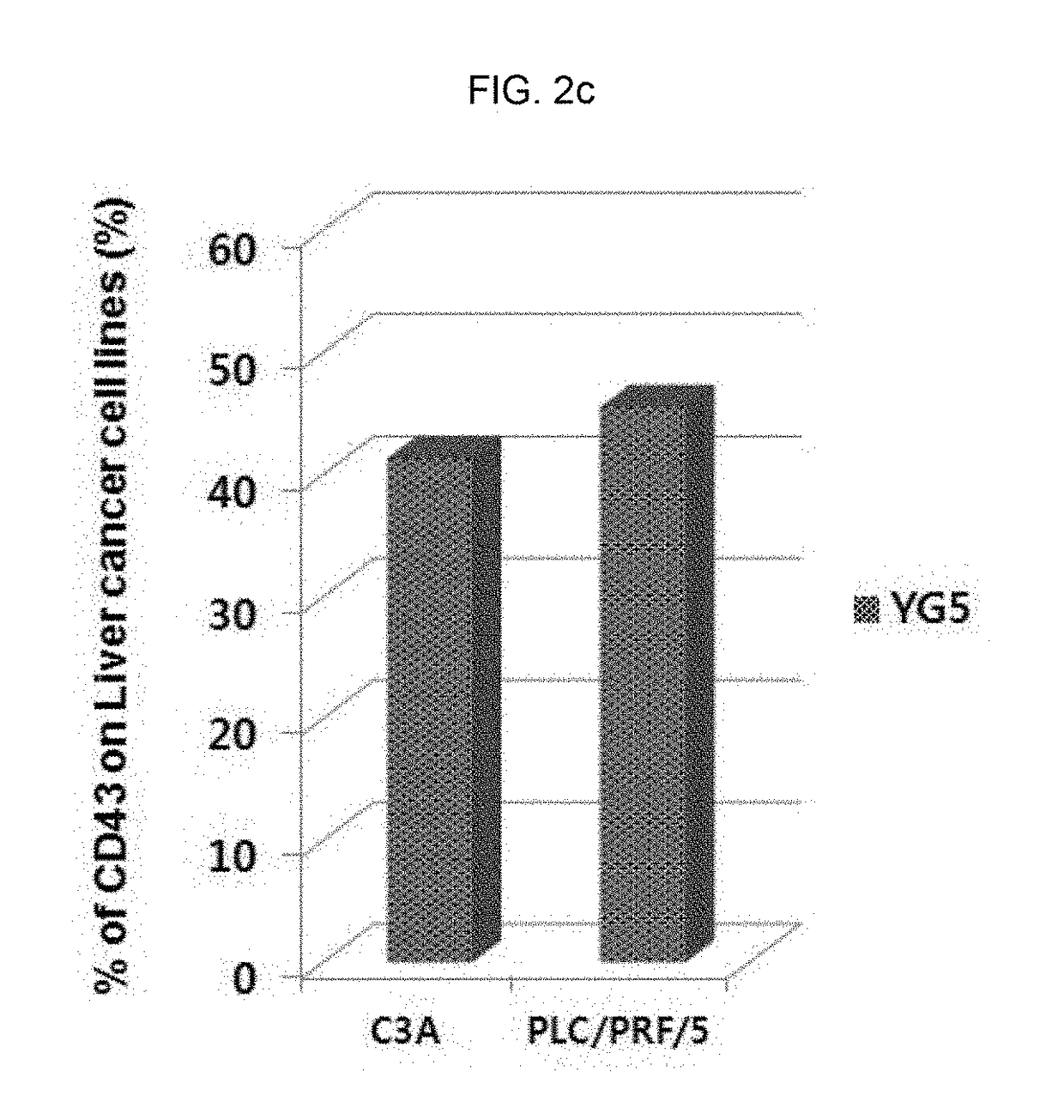
tion of Expression Level of CD43 Epitope in Human Solid Cancer Cell Lines
[0328]In order to investigate the expression level of CD43 in various solid cancer cell lines, immunostaining and flow cytometry were conducted.
[0329]The information of cell lines used for analysis was as follows:
NameOriginHistopathologyAccession NO.SNU-1stomach,adenocarcinomaATCC, CRL-5971gastricSNU-719stomachadenocarcinoma, primaryKOLB, No. 00719NCI-N87stomachcarcinoma; metastaticATCC, CRL-5822to liverAGSstomachadenocarcinomaATCC, CRL-1739HT29colonadenocarcinomaATCC, HTB-38LS174TcolonDukes' type B,ATCC, CL-188colorectaladenocarcinomaHCT116coloncolorectal carcinomaATCC, CCL-247C3Aliverhepatocellular carcinomaATCC, CRL-10741HepG2liverhepatoblastomaATCC, HB-8065PLC / PRF / 5liverhepatomaATCC, CRL-8024
[0330]Specifically, each cell line was inoculated and cultured in 100 mm of cell culture container, and when 70˜80% of surface was concentrated with the culture cell, the culture cell was washed with phosphate-buffered ...
example 3
ytotoxicity of Anti-CD43 Antibody to Cancer Cell (In Vitro)
[0332]3-1. Preparation of Antibody-Toxin Conjugate
[0333]The saporin (Sigma, St. Louis, Mo.) conjugation of monoclonal antibody was conducted according to the conventional method (Polito et al., 2004). After dissolving the antibody (DNP001; prepared in example 1-2) and saporin at the concentration of 2 mg / mL (antibody concentration) and 8 mg / mL (saporin concentration), respectively in 50 mM sodium borate buffer (pH 9.0), 2-iminothiolane (Sigma) was treated at the concentration of 0.4 mM and 1.0 mM, respectively. Afterward, the antibody and saporin were mixed at the ratio of 10:1 and reacted at the room temperature for 16 hours, and the antibody-saporin conjugate was purified by gel filtration. Hereinafter, the prepared conjugate was described as anti-CD43-saporin conjugate.
[0334]Referring the method above, anti-CD43-MMAE conjugate in which anti-CD43 antibody (DNP001; prepared in example 1-2) and monomethyl auristatin E (MMAE;...
PUM
| Property | Measurement | Unit |
|---|---|---|
| time | aaaaa | aaaaa |
| time | aaaaa | aaaaa |
| time | aaaaa | aaaaa |
Abstract
Description
Claims
Application Information