Laser speckle micro-rheology in characterization of biomechanical properties of tissues

a biomechanical property and micro-rheology technology, applied in the field of biological tissue mechanical properties measurement, can solve the problems of increasing the ecm stiffness, affecting the health of cells, and no means of 3d ecm stiffness measurement technology,

Active Publication Date: 2019-02-14
THE GENERAL HOSPITAL CORP
View PDF4 Cites 8 Cited by
  • Summary
  • Abstract
  • Description
  • Claims
  • Application Information

AI Technical Summary

Benefits of technology

[0023]In a related embodiment, the method additionally includes changing the optical distance by an incremental amount and repeating the steps of detecting, calculating, and associating as defined above in order to obtain and characterize a 2D distribution of the viscoelastic parameter corresponding to a layer of tissue located at a different, changed depth within the tissue sample. Generally, the incremental amount by which the optical distance is changed does not exceed 20 microns and is preferably smaller than 10 microns and more preferably smaller than 5 microns. In a specific implementation, changing the optical distance includes repositioning of an optical component of the sample train and adjusting a variable optical delay length by an amount optically matching said incremental amount. Detection of an optical interferogram optionally includes detection of a sample distribution of light that is defocused with respect to a boundary of the biological tissue, while changing the optical distance optionally includes adjusting the variable optical delay length. The method may additionally include displaying calculated 2D distributions as a 3D map of a geometrical distance parameter corresponding to the optical distance. The method may additionally include transforming 2D distributions calculated as a functions of depths of the tissue layers into the 3D map representing a 3D microscopically-resolved distribution of a mechanical property of the tissue.

Problems solved by technology

As a result, a positive feedback loop is established with consequences that are sometimes detrimental to the cell's health.
However, no means exists today that enable measurements of the ECM stiffness in 3D at microscopic size scales relevant to the microenvironment of a biological cell.
This, in turn, can accelerate the secretion, deposition, and cross linking of ECM macromolecules by host stromal cells, and results in further increases in ECM stiffness, completing a self-enforcing vicious cycle.
In addition, interference between cytoskeletal tension and the epidermal growth-factor receptor (EGFR) pathways can result in increased proliferation of cancer cells.
Further, increased ECM density and rigidity can amplify the interstitial fluid pressure and hamper drug delivery, promoting biophysical drug resistance.
One such disease is IPF, a devastatingly progressive fibrosis of the lungs that destroys the normal alveolar structures and impairs oxygen transfer to the blood stream.
Despite the ubiquitous role of ECM micro-mechanics in normal lung development and pulmonary disorders such as IPF, our current understanding of ECM mechanics is limited.
The limited cutaneous scleroderma affects only the skin on the face and limbs, whereas the diffuse type is likely to damage internal organs such as kidneys, heart, lungs, and digestive tract.
Currently, the cause of SS is unknown and no pharmacotherapy is available for this deadly disease.
The subsequent excessive deposition of ECM creates a feed-forward loop with catastrophic consequences.
Moreover, this type of mechanical test requires relatively large sample volumes and is not conducive for rare and precious biomaterials and tissue specimens.
Given that the approach-retract step takes at least a few seconds, the force-mapping AFM measurements are tediously time-consuming.
Moreover, due to their contact-based nature, the indentation-based technologies are inherently invasive.
In addition, the depth probed by the tip does not exceed a few microns, limiting AFM to a surface probe modality.

Method used


Image

  • Laser speckle micro-rheology in characterization of biomechanical properties of tissues
  • Laser speckle micro-rheology in characterization of biomechanical properties of tissues
  • Laser speckle micro-rheology in characterization of biomechanical properties of tissues

Examples


Embodiment Construction

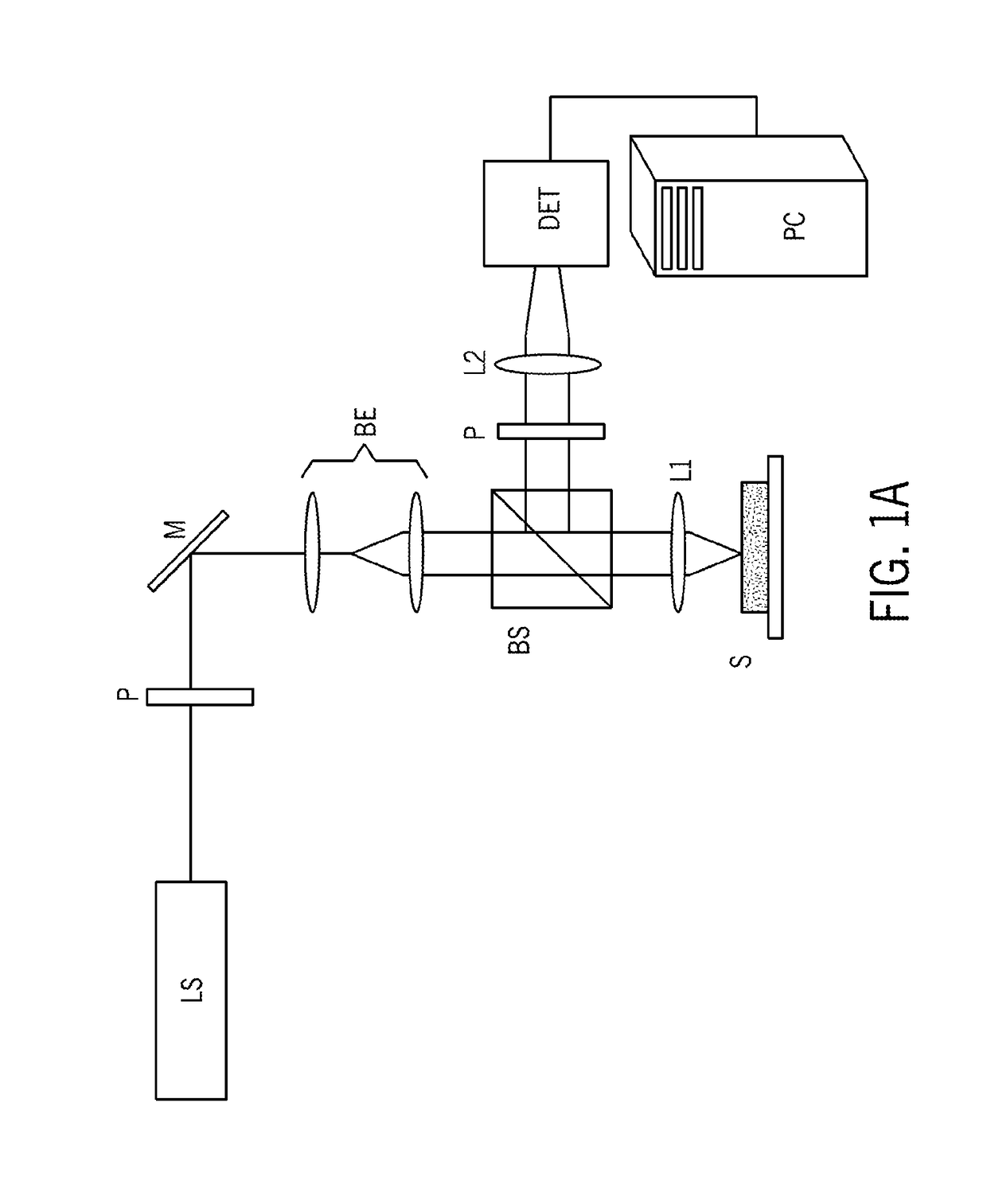
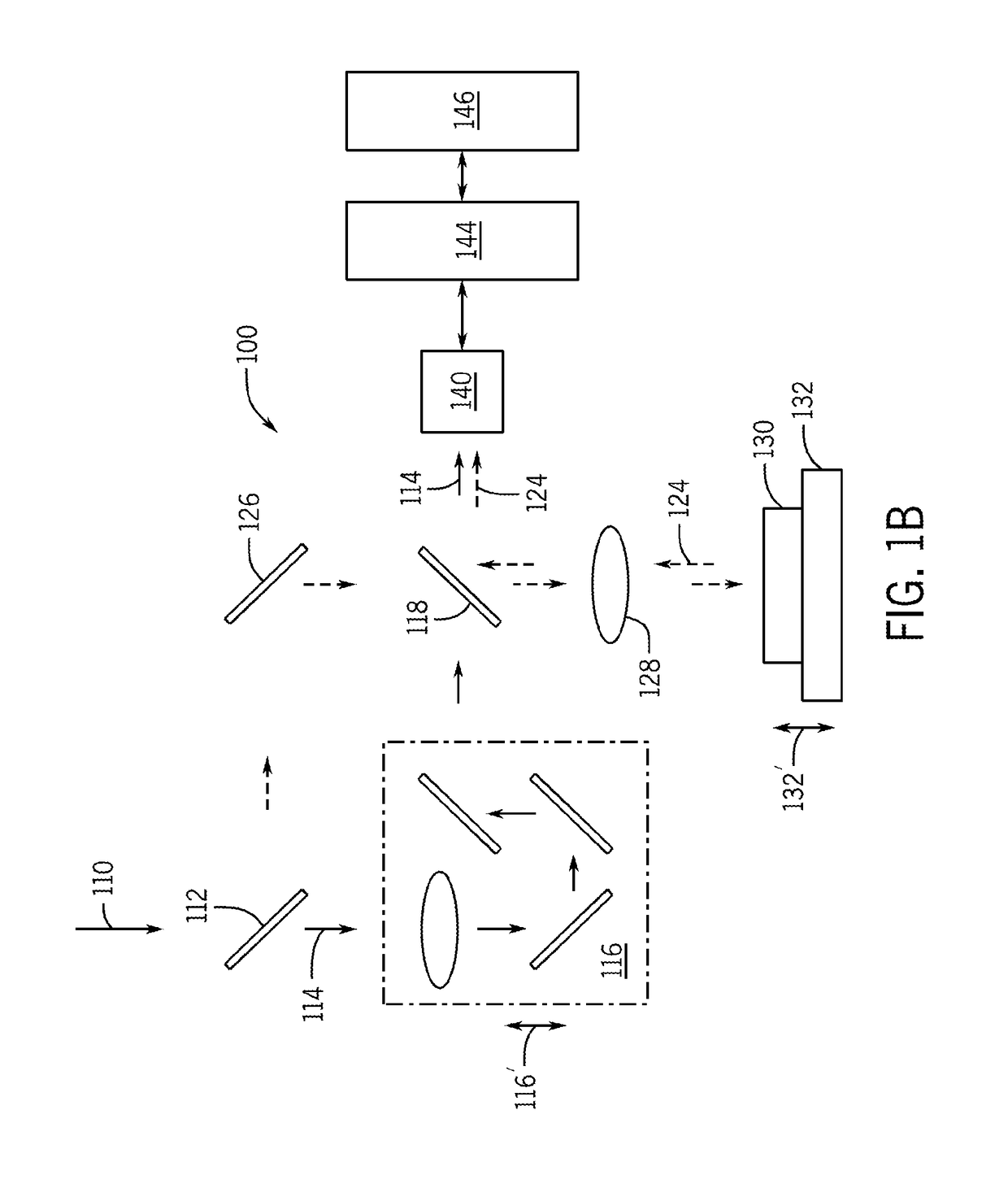
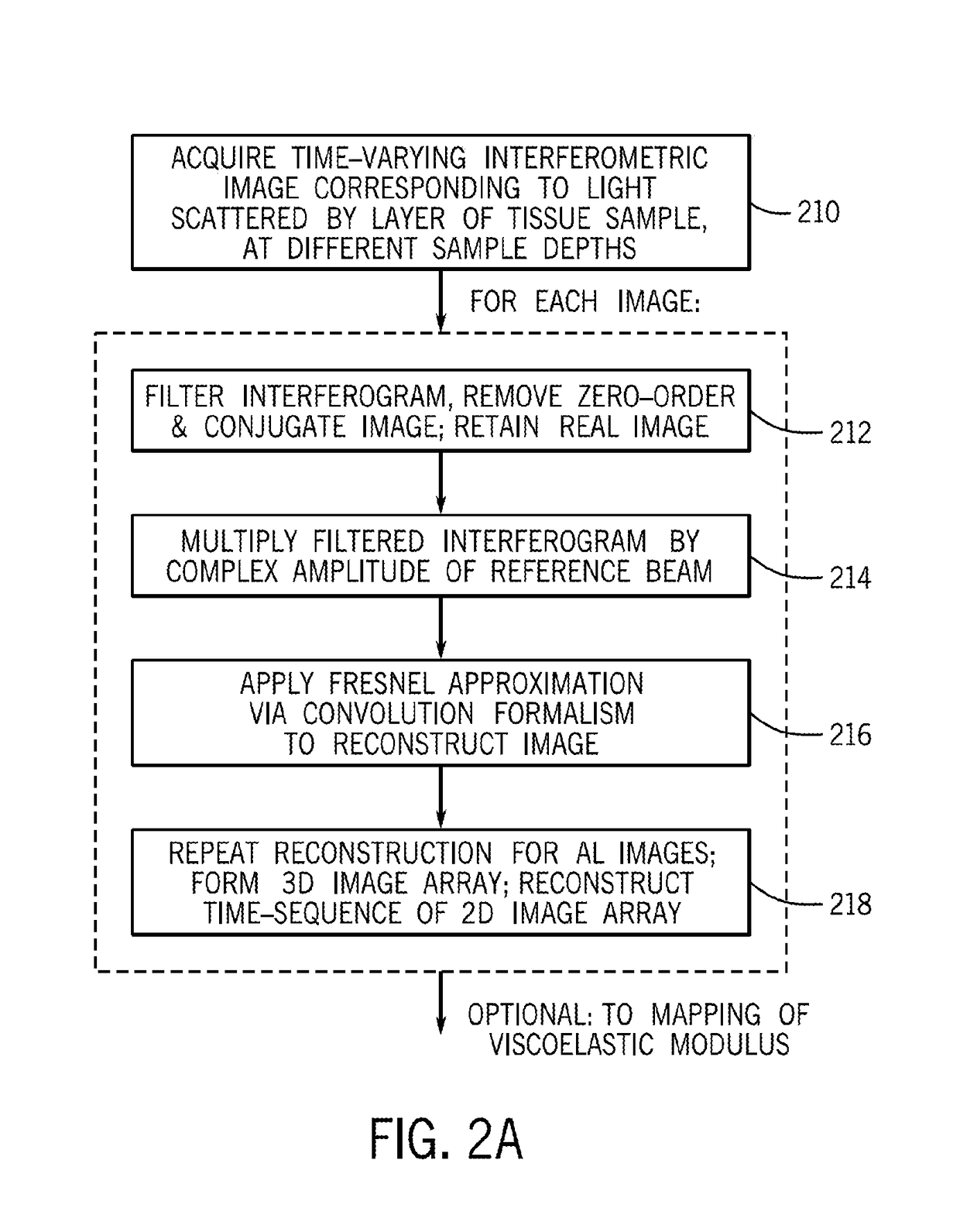
[0086]In accordance with preferred embodiments of the present invention, a Laser Speckle Microrheometer (LSM) system is disclosed, as well as multiple corresponding passive and active methods of depth-resolved speckle microrheometry, that facilitate the measurements of 3D mechanical properties of a biological tissue with cellular-scale (on the order of several microns, for example 1 to 20 microns, preferably 1 to 10 microns, and more preferably 1 to 5 microns) resolution with high sensitivity in order to monitor small changes in the ECM stiffness.

[0087]References throughout this specification to “one embodiment,”“an embodiment,”“a related embodiment,” or similar language mean that a particular feature, structure, or characteristic described in connection with the referred to “embodiment” is included in at least one embodiment of the present invention. Thus, appearances of the phrases “in one embodiment,”“in an embodiment,” and similar language throughout this specification may, but ...


PUM

PropertyMeasurementUnit
azimuth angleaaaaaaaaaa
optical distanceaaaaaaaaaa

Abstract

Laser speckle microrheology is used to determine a mechanical property of a biological tissue, namely, an elastic modulus. Speckle frames may be acquired by illuminating a coherent light and capturing back-scattered rays in parallel and cross-polarized states with respect to illumination. The speckle frames may be analyzed temporally to obtain diffuse reflectance profiles (DRPs) for the parallel-polarized and cross-polarized states. A scattering characteristic of particles in the biological tissue may be determined based on the DRPs, and a displacement characteristic may be determined based at least in part on a speckle intensity autocorrelation function and the scattering characteristic. A size characteristic of scattering particles may be determined based on the DRP for the parallel polarization state. The mechanical property may be calculated using the displacement and size characteristics.

Description

CROSS REFERENCE TO RELATED APPLICATIONS[0001]This application claims the benefit of U.S. Provisional Patent Application No. 62 / 294,453 filed Feb. 12, 2016, as well as U.S. Provisional Patent Application No. 62 / 420,948 filed Nov. 11, 2016. The disclosure of each of the above-cited applications, as well as the references cited therein, is hereby incorporated by reference.TECHNICAL FIELD[0002]The present invention relates to measurements of mechanical properties of a biological tissue and, more particularly, to measurements of such properties with cellular resolution with the use of a laser speckle microrheometer.BACKGROUND ART[0003]It is recognized that progression of such diseases as cancer and atherosclerosis, for example, and other debilitating disorders including neurodegenerative disease and osteoarthritis, is accompanied by changes in stiffness of biological tissue. Recent advances in the field of mechanobiology establish that these changes in the stiffness of the extra-cellular...

Claims


Application Information

Patent Timeline
Patent Type & Authority Applications(United States)
IPC IPC(8): G01N15/02G01N11/00G01N21/45G01N21/47G01N33/483G01N21/63
CPCG01N15/0227G01N11/00G01N21/45G01N21/4738G01N33/4833G01N21/636G01N2015/0065G01N2015/0233G01N2021/479G01N2021/4792G01N2203/0089
Inventor NADKARNI, SEEMANTINI K.HAJJARIAN, ZEINAB
Owner THE GENERAL HOSPITAL CORP
Why Eureka
Try Eureka