Systems and Methods for Identifying Protein Aggregates in Biotherapeutics

a biotherapeutics and protein technology, applied in the field of systems and methods for inspecting particles in liquid beneficial agents, can solve the problems of complex structure, difficult detection and control of molecular aggregation, and underlie great conformational flexibility and reduced or limited physical stability

Inactive Publication Date: 2019-05-23
ABBVIE INC
View PDF3 Cites 3 Cited by
  • Summary
  • Abstract
  • Description
  • Claims
  • Application Information

AI Technical Summary

Problems solved by technology

Protein biologics can present challenges different from other therapeutic solutions, such as small molecule drugs.
For example, and without limitation, protein biologics can be made from living cells and thus can have some degree of chemical and physical heterogeneity, and their molecular weight and structural complexity can underlie great conformational flexibility and reduced or limited physical stability.
The protein concentration in biopharmaceuticals can be high (for example and without limitation in a range of about 10-100 mg / mL), which can make molecular aggregation challenging to detect and control at all stages from production of the formulated protein through packaging and storage in the delivery device.
However, SEC can be considered a non-equilibrium invasive methodology.
As such, SEC can be unsuitable for investigating the dependence of aggregation on protein concentration, at least in part because SEC typically cannot be performed at the high protein concentrations relevant to the drug product, and substantial dilution (e.g., from the injected concentration) can occur during the chromatographic process.
The flow of aggregates larger than about 0.1 microns (e.g., column dependent) through columns may be restricted and thus can be excluded from detector chromatograms, which may result in a distorted analysis of aggregate size and extent.
However, each of these techniques can be considered invasive, can encounter procedural challenges when concentrations are moderately high, and may be unsuitable to distinguish protein particles from other particles, such as silicone oil (used as a lubricant in syringes), micro air bubbles, or other foreign particles.
However, these methods can be considered invasive, can have large sample volume requirements, in certain instances the assumptions may not be correct, and at least in part because image clarity can depend on the contrast afforded by particle refractive index and diffraction limited optical resolution, particle assignment can be increasingly difficult for particles below a certain size, such as below about 5 microns.
cur. In addition, there can be a large extrapolation factor intrinsic to the method (due at least in part to small fractional sampling), which can reduce the accuracy of quantitating components present at low particle number concentra
tion. As such, direct comparisons with the particle imaging methods can be difficult for samples with certain types of particles and particle size distribu
The invasive techniques described above, when used to evaluate prefilled syringes for quality control, can be unsuitable or impractical for evaluating more than a small fraction of syringes in a given manufacturing lot.
Certain commercial noninvasive inspection systems for prefilled syringes are available, but they can generally be used to detect only the presence of high concentrations of very large particles (e.g., exceeding about 25 microns), and can be unable to discriminate between particle types.

Method used


Image

  • Systems and Methods for Identifying Protein Aggregates in Biotherapeutics
  • Systems and Methods for Identifying Protein Aggregates in Biotherapeutics
  • Systems and Methods for Identifying Protein Aggregates in Biotherapeutics

Examples


Embodiment Construction

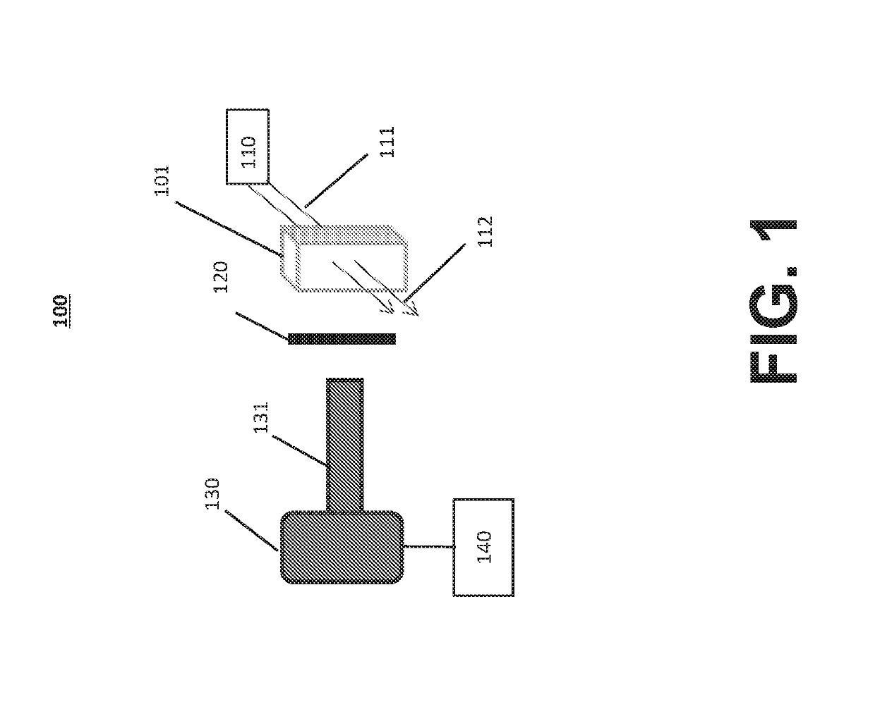
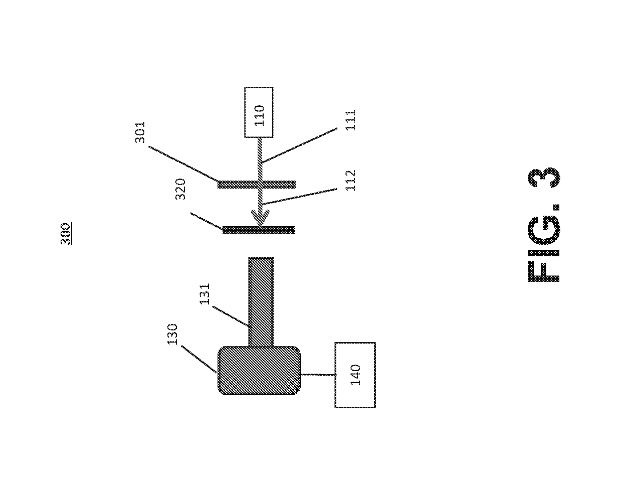
[0042]Reference will now be made in detail to the various exemplary embodiments of the disclosed subject matter, exemplary embodiments of which are illustrated in the accompanying drawings. The structure and corresponding method of operation of the disclosed subject matter will be described in conjunction with the detailed description of the system.

[0043]The systems and methods presented herein can be used for detection of particles, such as proteins and protein aggregates or any other visible or subvisible particles, in any of a variety of suitable beneficial agents or substances. As used herein, a “liquid beneficial agent” or “beneficial agent” (used interchangeably herein) is intended to refer generally to a substance or formulation in liquid form to be administered to or used by an individual (also referred to herein as a user or a patient) for an approved medical indication, such as a medication, diagnostic, nutritional, or other therapeutic agent. For example and without limit...


PUM

PropertyMeasurementUnit
concentrationaaaaaaaaaa
sizeaaaaaaaaaa
sizeaaaaaaaaaa

Abstract

Systems and methods for inspecting particles in a liquid beneficial agent are provided. Inspecting particles in a liquid beneficial agent includes selectively illuminating at least a portion of a liquid beneficial agent contained within a container using an excitation beam configured to excite photoluminescent particles in the liquid beneficial agent to emit an emission light and produce scattered excitation light, filtering the illuminated portion of the liquid beneficial agent to transmit the emission light and block the scattered excitation light, obtaining an image of the filtered emission light, analyzing image data representing the image of the filtered emission light to detect regions of the image representing the intrinsic photoluminescence of the photoluminescent particles, measuring an intensity of the regions of the image representing the intrinsic photoluminescence of the photoluminescent particles, and determining a size or number of the photoluminescent particles from the measured intensity of the regions.

Description

CROSS-REFERENCE TO RELATED APPLICATION[0001]This application claims the benefit of U.S. Provisional Patent Application No. 62 / 337,170, filed May 16, 2016, which is incorporated by reference herein in its entirety.BACKGROUNDField of the Disclosed Subject Matter[0002]The present disclosed subject matter relates to systems and methods for inspecting particles in a liquid beneficial agent, including systems and methods for identifying and distinguishing certain particles, such as protein monomers and aggregates, from other visible, subvisible, and submicron particles, such as silicone oil droplets and micro air bubbles, in a liquid beneficial agent, such as protein-based therapeutic solutions.Description of Related Art[0003]Protein biologics can present challenges different from other therapeutic solutions, such as small molecule drugs. For example, and without limitation, protein biologics can be made from living cells and thus can have some degree of chemical and physical heterogeneit...

Claims


Application Information

Patent Timeline
Patent Type & Authority Applications(United States)
IPC IPC(8): G01N15/02G01N15/06H04N5/225G06T7/60G06T7/00
CPCG01N15/0227G01N15/06H04N5/2256G06T7/60G06T7/0002G01N2015/0693G01N2015/0053G06T2207/10056G06T2207/30242G01N2015/0687G01N21/51G01N21/6456G01N21/6486G01N21/9027G01N21/53H04N23/56
Inventor MATAYOSHI, EDMUNDTU, SHENWANG, JIE
Owner ABBVIE INC
Why Eureka
Try Eureka