Methods for the treatment of cancer using meglumine
- Summary
- Abstract
- Description
- Claims
- Application Information
AI Technical Summary
Benefits of technology
Problems solved by technology
Method used
Image

Examples
example i
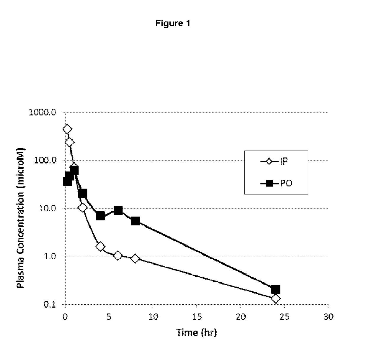
[0095]The pharmacokinetics of a single dose of meglumine hydrochloride was measured in male, HLA Swiss mice weighing 20-30 grams (FIG. 1). Animals (24 in each group) were dosed either by oral gavage (500 mg / kg in 0.25 ml) or intraperitoneally (100 mg / kg in 0.5 ml). A terminal brachial bleed of three mice was taken after 15 minutes, 30 minutes, 1 hour, 2 hour, 4 hour, 6 hour, 8 hour, and 24 hour of dosing. The pooled blood was collected into potassium EDTA coated tubes. Approximately 1 ml of blood was collected to obtain a 500 microliter sample of plasma. The tubes were spun for approximately 10 minutes at 8000 rpm, and plasma was drawn off into plastic tubes for storage at −80° C. prior to analysis.
[0096]Meglumine concentrations in plasma were determined using liquid chromatography-tandem mass spectrometry (LC-MS / MS). Plasma samples (25 microliters) were combined with 25 microliters of acetonitrile:water (1:1), and 25 microliters of 4 microgram / ml glucosamine as an internal standard...
example 2
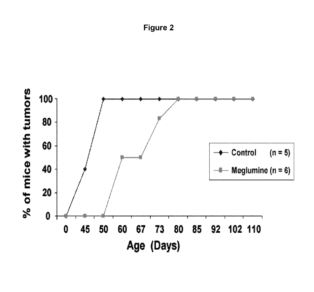
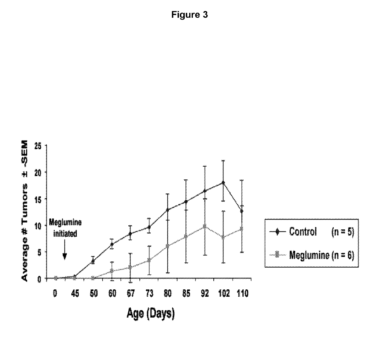
[0097]K6 / ODC transgenic mice are highly susceptible to developing skin tumors following a single treatment with a low subthreshold dose of the carcinogen DMBA. The effect of oral administration of meglumine in the drinking water was tested on the formation and growth of skin tumors in DMBA-initiated K6 / ODC transgenic mice and their normal littermates. Four day old K6 / ODC transgenic mice and their normal littermates were initiated with a single topical application of 300 nmol DMBA in 50 μl acetone. At birth the dam was given 0.5% DFMO in her drinking water to suppress ODC activity in K6 / ODC transgenic pups until weaned at 3 weeks of age when DFMO administration was stopped. DFMO has been shown to transfer to pups via the milk, and administration of this dose of DFMO has no adverse effects on the development of the mice.
[0098]Upon weaning, K6 / ODC transgenic mice and their normal littermates were divided into two treatment groups where they received ad lib either tap water or water wit...
example 3
[0102]THP-1 cells (human acute monocytic leukemia) were grown in a 24-well polystyrene plate in 1 ml of RPMI media with 10% fetal bovine serum. Cells were treated with 25 ng of lipopolysaccharide (LPS) (Sigma) in the presence or absence of 40 or 80 mM meglumine hydrochloride. After 24 hours, the media was centrifuged to remove the cells and analyzed for cytokine content using a Bio-Plex® immunoassay (Bio-Rad). The LPS induced levels of IL-17, IL-8, MIP-1 alpha, MIP-1 beta, IL-9 and IP-10 were decreased in the presence of meglumine (FIG. 5).
PUM
| Property | Measurement | Unit |
|---|---|---|
| Molar density | aaaaa | aaaaa |
| Dimensionless property | aaaaa | aaaaa |
| Frequency | aaaaa | aaaaa |
Abstract
Description
Claims
Application Information