Method and apparatus for characterizing an obstacle within an examination object using a medical image data set

Active Publication Date: 2019-06-06
SIEMENS HEALTHCARE GMBH
View PDF0 Cites 3 Cited by
  • Summary
  • Abstract
  • Description
  • Claims
  • Application Information

AI Technical Summary

Benefits of technology

[0047]One embodiment of the method provides that a further medical image data set is acquired according to the selected method. An advantage of this embodiment is that the properties supplied according to the inventive method can advantageously be used for a further acquisition. It can thereby be ensured that the examination object, in particular the examination region, comprising the obstacle can be displayed in an optimum quality and/or a quality desired by the user during acquisition of a further medical image data set. Repetition of acquisition of the further medical image data set can be avoided thereby. The overall length of examination of an examination object can be reduced and/or efficiently used thereby. This is convenient and/or cost-efficient.
[0048]In another embodiment of the method, the medical image data set and the further medical image data set are acquired with an identical medical imaging apparatus (scanner). According to the inventive method, the original medical image data set can have been ac

Problems solved by technology

Obstacles of this kind typically influence medical imaging and can generate artifacts in the corresponding image data.
In the case of computed tomography systems having an elevated radiation dose, and in the case of magnetic resonance apparatuses the

Method used


Image

  • Method and apparatus for characterizing an obstacle within an examination object using a medical image data set
  • Method and apparatus for characterizing an obstacle within an examination object using a medical image data set

Examples


first embodiment

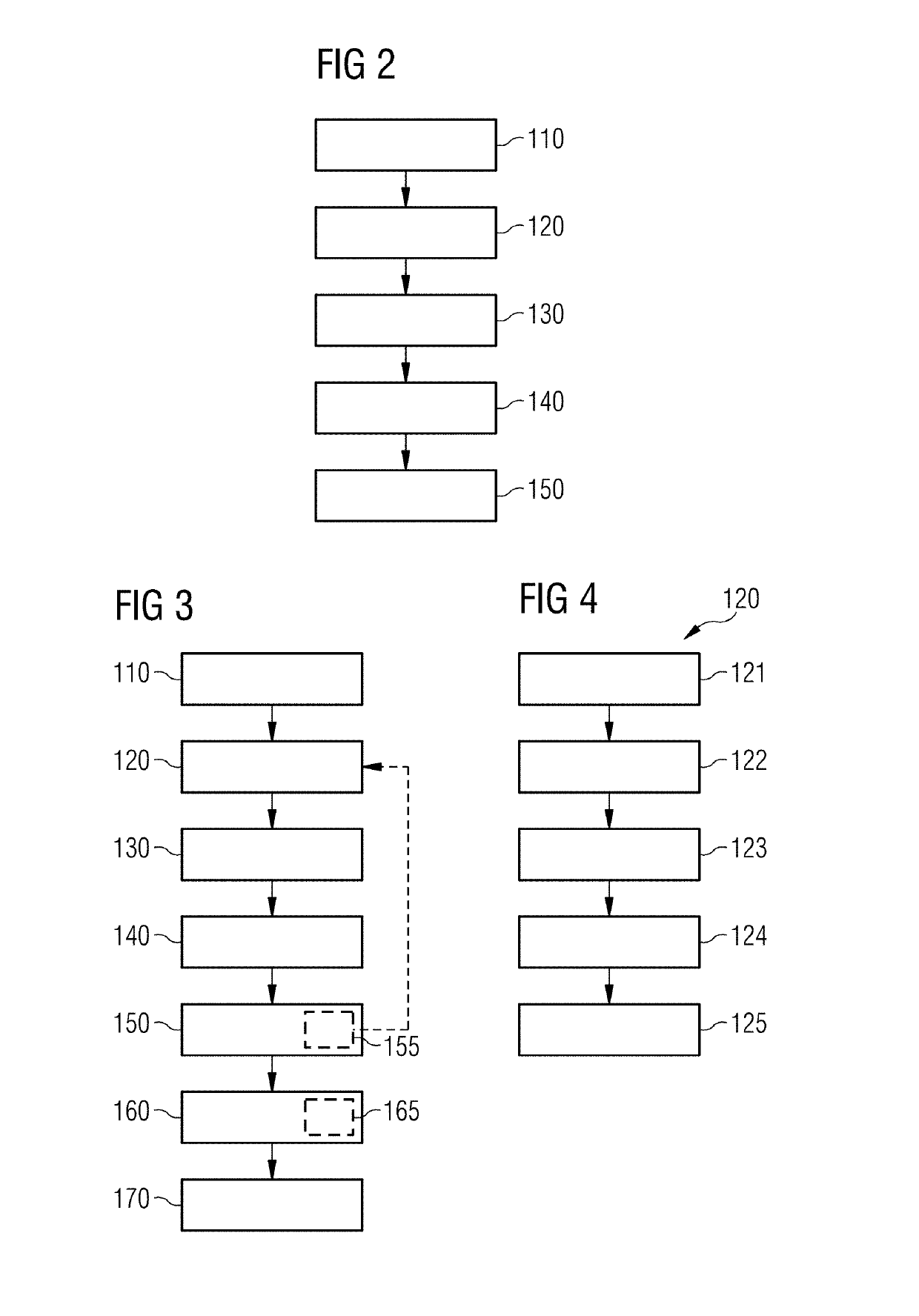
[0094]FIG. 2 is a flowchart of the inventive method. In method step 110 a database is supplied, comprising at least one data class for a feature for characterizing different obstacles each having at least one data entry for one obstacle respectively. In the following method step 120 a trained artificial neural network is supplied for relationships between the different obstacles, the feature and the medical image data set. In method step 130 the medical image data set is acquired from the examination object comprising the obstacle within the examination object. In method step 140 a property of the obstacle within the examination object is determined by applying the trained artificial neural network to the medical image data set, with a data entry of the data class being assigned to the obstacle within the examination object and the obstacle within the examination object being characterized by the data entry. In method step 150 the property of the obstacle within the examination obje...

second embodiment

[0096]FIG. 3 is a flowchart of an inventive method. The property of the obstacle within the examination object can be supplied according to method step 150 in such a way that, based thereon, a method for acquisition of a further medical image data set is selected. The selection can optionally comprise method step 165: an optimization of a method for acquisition of a further medical image data set. A criterion specified by a user can be considered for this. In method step 170 a further medical image data set is acquired according to the selected method. The further medical image data set is preferably acquired with the same medical imaging apparatus 12 as the original acquisition of the medical image data set.

[0097]If the medical imaging apparatus 12 is designed as a magnetic resonance apparatus 11, the further medical image data set is preferably acquired in method step 170 according to one of following magnetic resonance control sequences and / or recording techniques: TSE, SEMAC, VA...


PUM


Abstract

In a method and apparatus for characterizing an obstacle within an examination object using a medical image data set by the use of a database containing at least one data class, a trained artificial neural network defines and develops relationships between different obstacles, features and medical imaging data sets. A data entry of a data class is assigned by the neural network to the obstacle within the examination object and the obstacle within the examination object is characterized in an electronic output by this data entry.

Description

BACKGROUND OF THE INVENTIONField of the Invention[0001]The invention concerns a method, a medical imaging apparatus, a computer, and a non-transitory, electronically readable data carrier, for characterizing an obstacle within an examination object.Description of the Prior Art[0002]Medical imaging apparatuses, for example a magnetic resonance apparatus, a Single Photon Emission Computed Tomography (SPECT) apparatus, a Positron Emission Tomography (PET) apparatus, a computed tomography (CT) apparatus, an ultrasound device, an X-ray apparatus, a C-arm device, or a combined medical imaging apparatus, formed by any combination of such apparatus are suitable for generating a medical image data set.[0003]In a magnetic resonance apparatus, the body of an examination object to be examined, such as a patient, is conventionally exposed to a strong basic magnetic field, for example of 1.5 or 3 or 7 tesla, produced by a basic field magnet. In addition, gradient pulses are activated by a gradien...

Claims


Application Information

Patent Timeline
IPC IPC(8): A61B5/00A61B5/055G01R33/54G01R33/56
CPCA61B5/7267A61B5/055G01R33/543G01R33/5608G16H50/70
Inventor PAUL, DOMINIKZELLER, MARIO
Owner SIEMENS HEALTHCARE GMBH
Why Eureka
Try Eureka