Exosomes and mirna to treat glaucoma

a technology of mirna and exosomes, applied in the field of glaucoma, can solve the problems of severe detriment to vision, no neuroprotective strategy currently exists, and dysfunction and subsequent loss, and achieve the effect of increasing the survival rate of retinal ganglion cells

Active Publication Date: 2019-07-25
UNITED STATES OF AMERICA
View PDF0 Cites 6 Cited by
  • Summary
  • Abstract
  • Description
  • Claims
  • Application Information

AI Technical Summary

Benefits of technology

[0005]Methods are disclosed herein for treating glaucoma in a subject. In some embodiments, the methods increase retinal ganglion cell survival.

Problems solved by technology

RGC are irreplaceable, making their dysfunction and subsequent loss a severe detriment to vision and thus, quality of life.
While current therapies can successfully reduce intraocular pressure (IOP), the critical risk factor associated with glaucoma, no neuroprotective strategies currently exist.

Method used


Image

  • Exosomes and mirna to treat glaucoma
  • Exosomes and mirna to treat glaucoma
  • Exosomes and mirna to treat glaucoma

Examples


example 1

Methods

[0178]Animals:

[0179]Adult female Sprague-Dawley rats weighing 150-200 g (Charles River, Wilmington, Mass.) were maintained in accordance with guidelines described in the ARVO Statement for the Use of Animals in Ophthalmic and Vision Research, using protocols approved by the National Eye Institute Committee on the Use and Care of Animals.

[0180]Animals were kept at 21° C. and 55% humidity under a 12 hours light and dark cycle, given food / water ad libitum and were under constant supervision from trained staff. Animals were euthanized by rising concentrations of CO2 before extraction of retinae.

[0181]Materials:

[0182]All reagents were purchased from Sigma (Allentown, Pa.) unless otherwise specified.

[0183]BMSC Cultures:

[0184]Human CD29+ / CD44+ / CD73+ / CD90+ / CD45− BMSC (confirmed by supplier; Lonza, Walkersville, Md.) from 3 donors were pooled and cultured in DMEM containing 1% penicillin / streptomycin and 10% exosome-depleted foetal bovine serum (Thermo Fisher Scientific, Cincinnati, O...

example 2

BMSC Secreted Exosomes

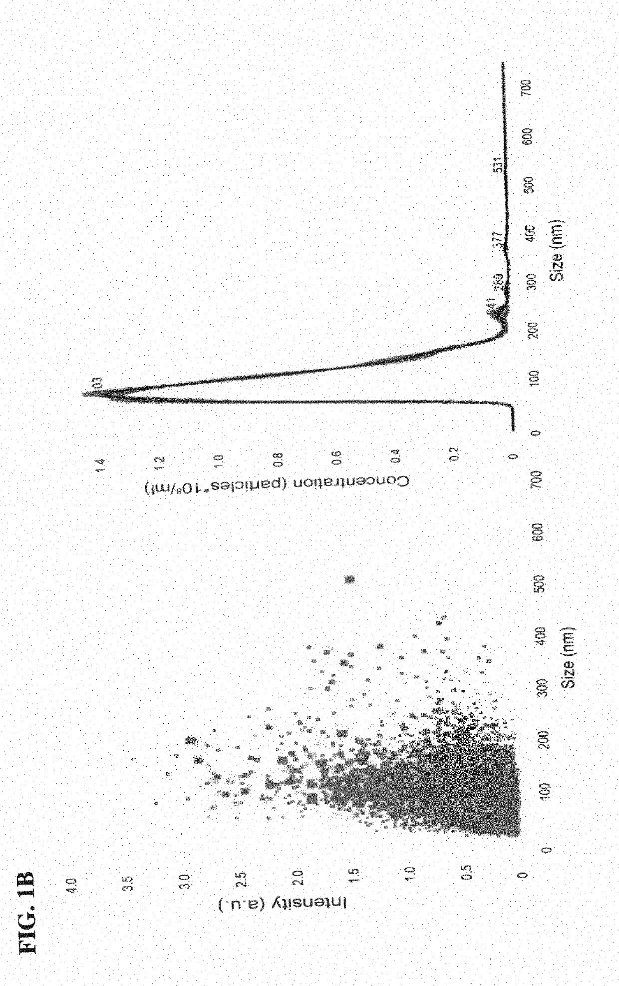
[0213]Exosomes isolated from human BMSC and fibroblasts were visualized clearly by NANOSIGHT™ and had a diameter of 100-120 nm, as expected for exosomes (FIG. 1B). Very few larger extracellular vesicles were detected and many of those that were detected were likely exosomal aggregates. No exosomes below 100 nm were detected due to limitations of the NANOSIGHT™ technology.

example 3

Intracameral Microbeads and Laser Photocoagulation of the Trabecular Meshwork LED to Elevations in IOP

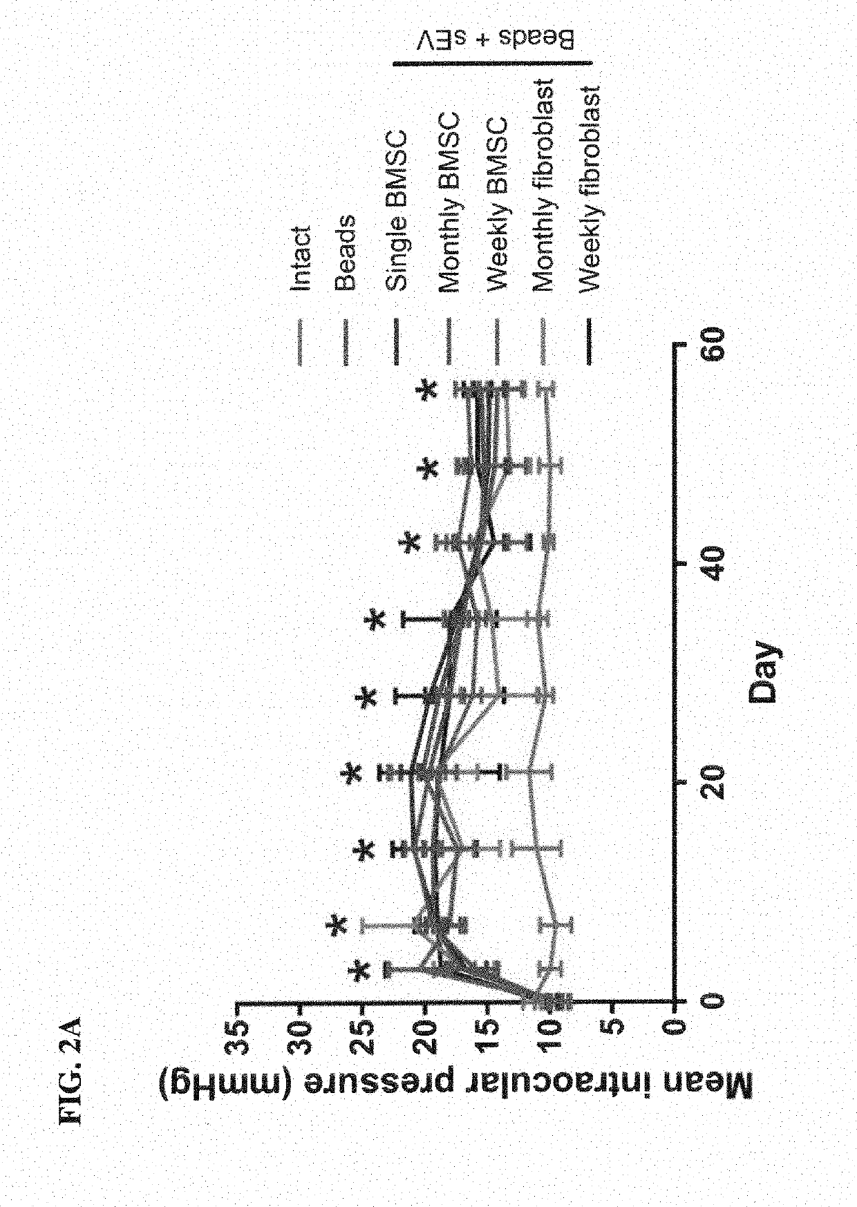
[0214]Microbeads delivered ic (Group 2) led to a significant rise in IOP from 9.0±0.5 mmHg (Day 0) to 20.5±2.4 mmHg (Day 3), which remained high till the end of the experiment (14.2±2.0 mmHg; Day 56; FIG. 2A). In contrast, IOP in uninjected eyes (Group 1; 11.4±0.7 mmHg; Day 0) did not change significantly (10.3±0.6 mmHg; Day 56). The injection of exosomes into the vitreous did not significantly affect the IOP in ic microbead injected eyes.

[0215]Laser photocoagulation (Group 3) led to a significant rise in IOP from 9.0±0.4 mmHg (Day 0) to 25.0±5.8 mmHg (Day 7), which remained high till Day 17 (20.1±4.2 mmHg) but returned to baseline by the end of the experiment (11.1±1.4 mmHg; Day 21; FIG. 2B). In contrast, IOP in uninjected eyes (Group 1; 11.4±0.7 mmHg; Day 0) did not change significantly (11.7±1.8 mmHg; Day 21). The injection of exosomes into the vitreous did not significantly affe...


PUM

PropertyMeasurementUnit
distancesaaaaaaaaaa
distancesaaaaaaaaaa
distancesaaaaaaaaaa

Abstract

Methods are disclosed herein for treating glaucoma in a subject. In some embodiments, the methods increase retinal ganglion cell survival. The disclosed method use exosomes and/or miRNA.

Description

CROSS REFERENCE TO RELATED APPLICATIONS[0001]This claims the benefit of U.S. Provisional Application No. 62 / 622,032, filed Jan. 25, 2018, which is incorporated herein by reference.STATEMENT OF GOVERNMENT SUPPORT[0002]This invention was made with Government support under project number Z01#: EY000318-20 by the National Institutes of Health, National Eye Institute. The Government has certain rights in the invention.FIELD OF THE DISCLOSURE[0003]This relates to the field of glaucoma, specifically methods for treating glaucoma using exosomes or a vector encoding a miRNA.BACKGROUND[0004]Glaucoma is one of the world's leading causes of irreversible blindness and is characterized by the slow progressive degeneration of retinal ganglion cells (RGC) and their axons (Tham et al., Ophthalmology 2014; 121:2081-2090). RGC operate as the final stage in the phototransductive visual pathway of the retina, tasked with the projection of electrochemical information to the brain along their axons which ...

Claims


Application Information

Patent Timeline
Patent Type & Authority Applications(United States)
IPC IPC(8): A61K35/28A61P27/10C12N5/0775A61K9/00C12N15/113
CPCA61K35/28A61P27/10C12N5/0663A61K9/0048A61K9/0019C12N15/113A61K45/06C12N2310/14C12N2310/141C12N2501/65A61K31/4178A61K31/5575A61K31/5377A61K31/498A61K2300/00C12N2320/30
Inventor TOMAREV, STANISLAV IVANOVICHMEAD, BENJAMIN FRANK JOHN MARTIN
Owner UNITED STATES OF AMERICA
Why Eureka
Try Eureka