Freeze-dried fibrin matrices and methods for preparation thereof

a fibrin matrix and freeze-dried technology, applied in the field of freeze-dried biomatrices, can solve the problems of short-lived clinical improvement, reduced function, damage to cartilage, etc., and achieve the effects of improving cell incorporation capacity, improving cell absorption ability, and maintaining viability

Inactive Publication Date: 2006-08-03
PROCHON BIOTECH
View PDF4 Cites 43 Cited by
  • Summary
  • Abstract
  • Description
  • Claims
  • Application Information

AI Technical Summary

Benefits of technology

[0212] One particular advantage of the present invention is the high level of cell viability and excellent cell distribution following cell seeding directly on a dry sponge. Often a matrix comprising an exogenous anti-fibrinolytic agent such as tranexamic acid exhibits lower cell viability following seeding. The cells seem to recover but the exogenous anti-fibrinolytic agents may be detrimental to initial cell growth. When such a sponge is washed and some or all of the tranexamic acid is removed cell proliferation is improved. It is also noted that many cells settle at the periphery of the matrix following on a wet sponge while there is a better cell distribution following seeding on a dry sponge.
[0213] Sponges comprising different concentrations of plasma proteins and thrombin were tested. Sponges comprising 10 mg / ml, 15, 16.5 mg / ml, 18 mg / ml, 20 mg / ml, 22 mg / ml, 25 mg / ml, 30 mg / ml and varying concentrations of hyaluronic acid (from about 0.05% to about 1.1%) and either 1, 1.5 or 2 IU thrombin / mg proteins. A total of about 5×10⁵ to about 5×10⁶ chondrocytes were seeded on 1 cm diameter sponges and allowed to incubate for three days. Different volumes of growth media were added and the cell-embedded matrix allowed to incubate. It is to be understood that the sponge of may be of varying sizes, shapes and thickness.
[0214] Following a three-day, 1 week and three week incubation for the seeded sponges, some of the sponges were collagenase degraded and cells counted following trypan blue staining. Cell proliferation is determined as described in Example 8, below.
[0215] Samples of the cell-bearing sponges or matrices, were paraffin-embedded and sections prepared using a microtome. The histological sections are further stained using different biological stains including hematoxylin and eosin (H&E), toluidine blue and fast red, Masson's trichrome stain and others. All sponges exhibited similar cell distribution, with live cells present throughout all layers of the sponge.
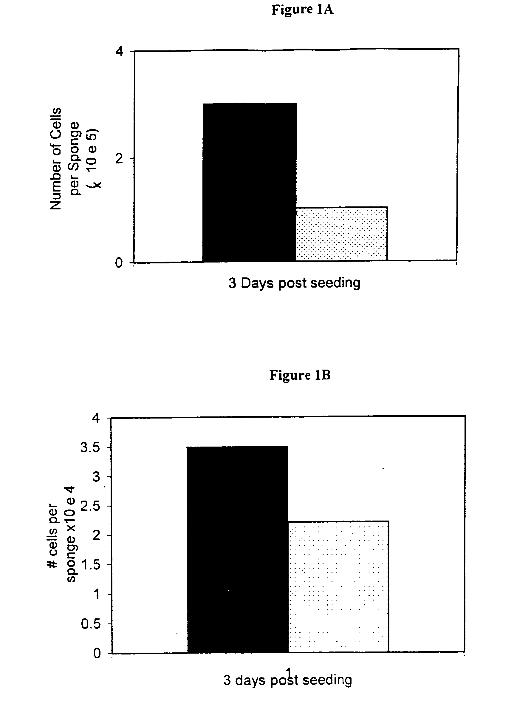
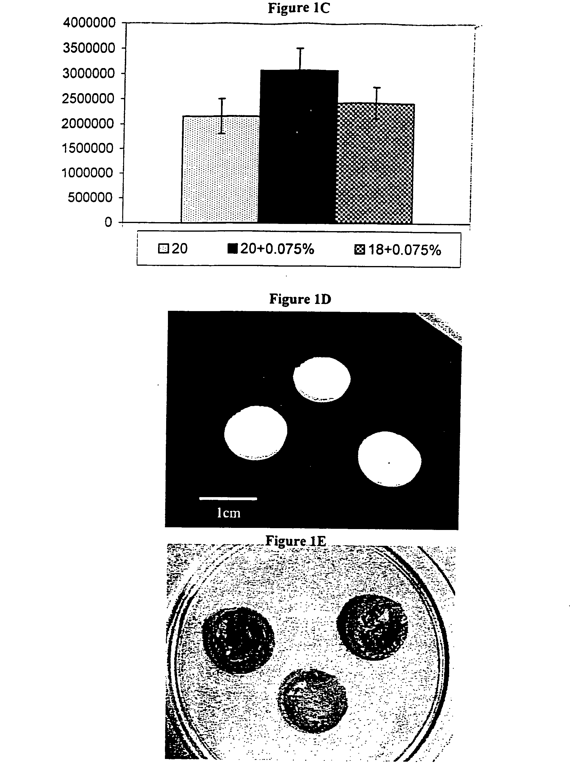
[0216] Examples of cell growth in the fibrin sponge of the invention are shown in FIGS. 1A and 1B and FIG. 2. Each sponge was seeded with 5×10⁶ porcine or human chondrocytes in 30 microliter volume, allowed to incubate one hour and fresh media was added. After three days the sponges were degraded in collagenase and the number of live cells was counted after staining with trypan blue. FIG. 1A shows the increased viability of porcine chondrocytes following a three day incubation seeded on matrices with (speckled) and without plasminogen (solid). After three days, more than 50% of the cells remained viable as compared to about 20% of the cells seeded on the standard matrix prepared from plasma proteins comprising tranexamic acid. FIG. 1B shows the viability of human chondrocytes seeded on matrices with (speckled) and without plasminogen (solid-dry) following a three-day incubation. The plasminogen free sponges showed superior cell viability when seeded with human chondrocytes when compared to the sponges comprising tranexamic acid. FIG. 1C shows cell viability after three days on three different sponge compositions. All sponges comprised plasma proteins substantially devoid of plasminogen and were seeded with about 4×10⁶ human chondrocytes. The speckled bar represents viability on a 20 mg plasma protein / ml sponge, without additive present. The solid black bar represents cell viability on a sponge comprising 20 mg / ml and 0.075% hyaluronic acid. The checkered bar represents cell viability on a sponge comprising 18 mg / ml and 0.75% hyaluronic acid. It can be seen that all three sponges provide a good scaffold for cell seeding. FIGS. 1D and 1E show photographs of one centimeter (1 cm) diameter dry sponges and as cell-bearing implants, respectively.
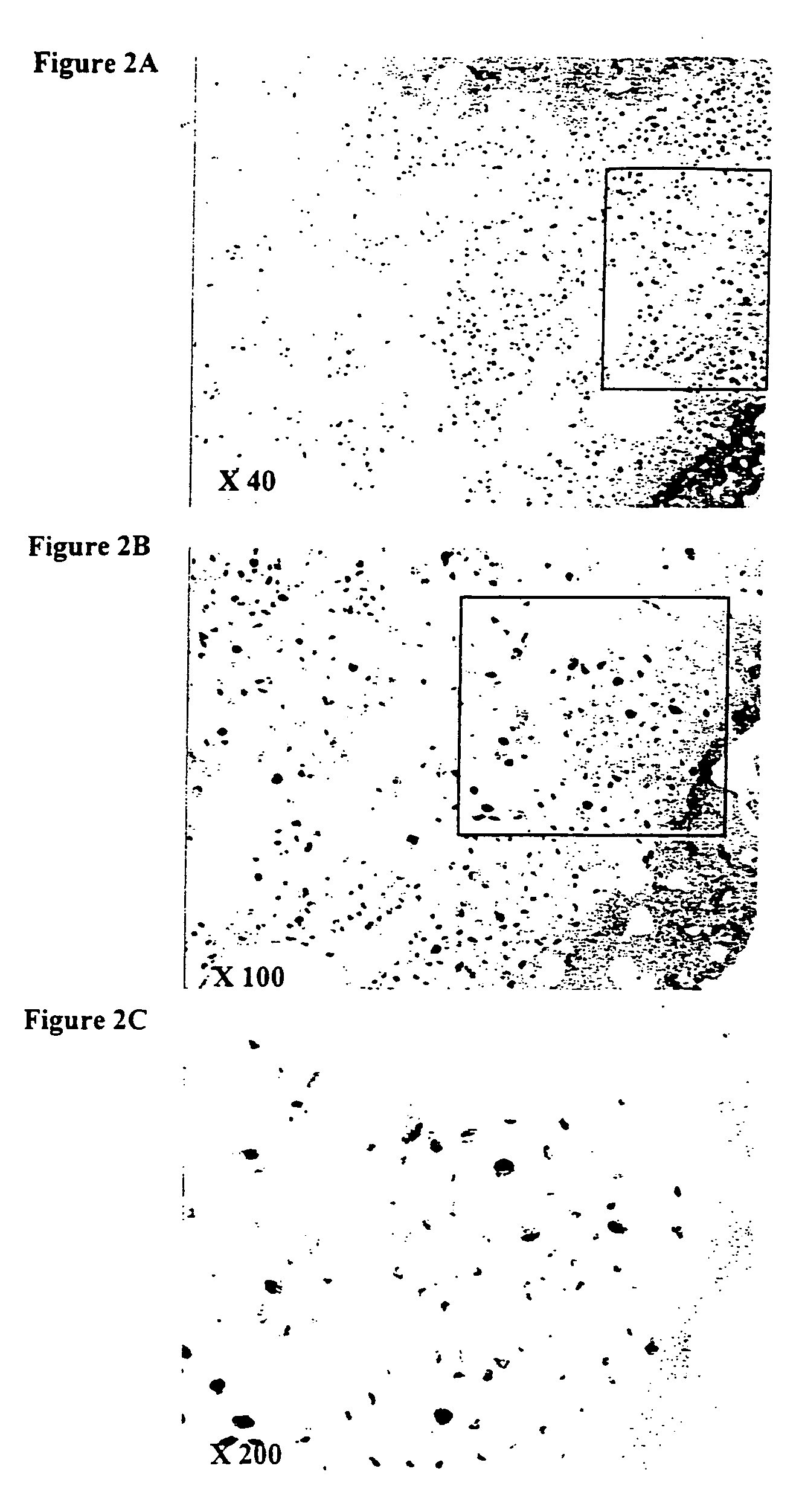
[0217]FIG. 2A-2C show histological cross sections through the center of a matrix comprising human chondrocytes following 1-week incubation. The fibrin sponge was made of commercial fibrinogen substantially devoid of plasminogen (Omrix, 20 mg / ml) comprising 0.05% hyaluronic acid and 1×10⁶ human cells. Note the infiltration of the chondrocytes into the sponge. FIGS. 2A, 2B and 2C show 40×, 100× and 400× magnifications, respectively. Example 7 in vitro Degradation Assay

Problems solved by technology

Damage to cartilage may result from an inflammatory disease such as rheumatoid arthritis, from a degenerative process such as osteoarthritis or from trauma such as intraarticular fracture or following ligament injuries.
Cartilage lesions are often associated with pain and reduced function, generally do not heal and without medical intervention may require total joint replacement.
Often the induced tissue is of a mixed fibrocartilage type, is not durable and the clinical improvements are short lived.
In fact, this technique requires a complicated procedure involving two surgical sites, and shows high variability and limited clinical success.
Existing freeze-dried fibrin implants for tissue engineering purposes are prepared using fibrinogen or plasma protein solutions having inherent proteases that may compromise the stability of certain of the plasma proteins and lead to degradation of the matrix.
These agents may have detrimental effects on cell growth, proliferation and / or differentiation or may cause adverse reactions in patients.
The art has not heretofore provided a stable freeze-dried fibrin matrix substantially devoid of exogenous anti-fibrinolytic agents.

Method used


Image

  • Freeze-dried fibrin matrices and methods for preparation thereof
  • Freeze-dried fibrin matrices and methods for preparation thereof
  • Freeze-dried fibrin matrices and methods for preparation thereof

Examples


example 1

Preparation of a Fibrin Matrix

[0176] Although detailed methods are given for the preparation of the plasma protein, it is to be understood that other methods of preparing plasma proteins are known in the art and are useful in the preparation of the matrix of the present invention. A non-limiting example of a protocol for the preparation of a fibrinogen-enriched solution is given in Sims, et al. (Plastic & Recon. Surg. 101:1580-85, 1998). Any source of plasma proteins may be used, provided that the plasma proteins are processed to be substantially devoid of anti-fibrinolytic agents, plasminogen and of organic chelating agents Examples of plasma protein preparation methods are given in examples 2 and 3, hereinbelow.

[0177] Materials and Methods:

[0178] Source of plasma proteins e.g. Plasminogen-free fibrinogen (Omrix, Ill.) approximately 50-65 mg / ml stock solution.

[0179] Calcium Chloride 5 mM

[0180] Thrombin (1000 International Units / ml, Omrix, Ill.)

[0181] Optional: Hyaluronic acid...

example 2

Isolation of Partially Purified Plasma Proteins from Whole Plasma

[0187] Plasma protein may be prepared from different sources such as fresh plasma, fresh frozen plasma, recombinant proteins and xenogeneic, allogeneic or autologous blood. The fresh frozen plasma was received from the blood bank (Tel-Hashomer, Israel). The plasma (220 ml) was thawed in a 4° C. incubator over night, followed by centrifugation at 4° C. at approximately 1900 g for 30 min. The pellet was resuspended in 2.5 ml PBS with gentle rolling until a homogenized solution was seen. The total protein concentration may be estimated by Bradford assay and SDS-PAGE (comparing to a standard). Exemplary samples were found to be about 42 mg total protein / ml to about 50 mg total protein / ml. The plasma may further be treated to remove plasminogen, using methods known in the art. Non-limiting examples of methods useful for removing plasminogen from blood or blood derivates such as plasma or a cryoprecipitate are disclosed in ...

example 3

Extraction of Plasma Protein Fractions from Allogeneic or Autologous Blood

Materials:

[0190] 1) Sodium citrate, 3.8% or any other pharmaceutically acceptable anti-coagulant [0191] 2) Ammonium sulfate (NH4)2SO4, saturated (500 g / l) [0192] 3) Ammonium sulfate (NH4)2SO4, 25% [0193] 4) Phosphate-EDTA buffer: 50 mM phosphate, 10 mM EDTA, pH 6.6 [0194] 5) Tris-NaCl buffer: 50 mM Tris, 150 mM NaCl, pH 7.4 [0195] 6) Ethanol, absolute 4° C. [0196] 7) Whole blood (Israel Blood Bank, Tel Hashomer Hospital or from patient)

Methods:

[0197] This method may be used to produce plasma proteins that may be treated for removal of plasminogen by methods known in the art, including affinity chromatography. The plasma proteins are isolated according to standard methods. To one 450 ml bag of blood from the blood bank, containing sodium citrate, 50 ml of a 3.8% sodium citrate solution was added and the solution was mixed gently.

[0198] The blood-sodium citrate was centrifuged at 2,100 g for 20 min. The s...


PUM

PropertyMeasurementUnit
concentrationaaaaaaaaaa

Abstract

The present invention relates to porous freeze-dried fibrin matrices substantially devoid of external anti-fibrinolytic agents, and methods of producing such matrices. Resilient matrices, also known as sponges, that are particularly beneficial for supporting three dimensional cell growth are obtained from plasma proteins substantially devoid of plasminogen or from partially purified plasma proteins, thus obviating the need for exogenous anti-fibrinolytic agents. Furthermore, incorporation of glycosaminoglycans and bioactive agents during the formation of the matrix results in a sponge having advantageous biological, mechanical and physical properties. The compositions of the present invention are useful clinically, per se or as cell-bearing implants.

Description

CROSS-REFERENCE TO RELATED APPLICATIONS [0001] This application is a continuation of International application PCT / IL2004 / 000088 filed Jan. 29, 2004, and claims the benefit of provisional application 60 / 507,167filed Oct. 1, 2003, the entire content of each of which is expressly incorporated herein by reference thereto.FIELD OF THE INVENTION [0002] The present invention concerns freeze-dried biomatrices comprising plasma proteins substantially devoid of plasminogen useful for clinical applications including as implants for tissue engineering. Additional preferred embodiments of the invention are freeze-dried plasma protein biomatrices substantially devoid of exogenous anti-fibrinolytic agents. The matrices according to the present invention are useful clinically, per se or as cell-bearing implants. BACKGROUND OF THE INVENTION [0003] Tissue Engineering. Tissue engineering may be defined as the art of reconstructing or regenerating mammalian tissues, both structurally and functionally ...

Claims


Application Information

Patent Timeline
Patent Type & Authority Applications(United States)
IPC IPC(8): A61K38/37A61K38/18A61K31/727A61K31/728A61K9/14A61K9/00A61K9/20A61K35/12A61L27/22A61L27/38A61L27/56C07K14/75C12N5/00C12N5/077
CPCA61F2/30756A61K9/0024A61K9/2063A61K31/727A61K31/728A61K35/12A61L27/225A61L27/3852A61L27/56C07K14/75C12N5/0068C12N5/0655C12N2533/56A61K38/1825A61L27/3804A61L27/3834A61P17/02A61P19/04
Inventor YAYON, AVNERAZACHI, MALKITGLADNIKOFF, MICHA
Owner PROCHON BIOTECH
Why Eureka
Try Eureka