Quantitative Shear Wave Elasticity Imaging Method and System

a quantitative and elastic imaging technology, applied in the field of medical ultrasound imaging, can solve the problems of difficult to reproduce detection, difficult to obtain quantitative detection information for disease tracking and postoperative observation, and difficult to directly compare, so as to achieve better reliability, stronger anti-noise capability, and better reliability

Active Publication Date: 2019-08-22
SASET CHENGDU TECH LTD
View PDF0 Cites 2 Cited by
  • Summary
  • Abstract
  • Description
  • Claims
  • Application Information

AI Technical Summary

Benefits of technology

[0041]The ultrasound quantitative elasticity imaging method and system provided in the present invention are based on a sliding window linear fitting strain and use a two-dimensional linear fitting shear wave velocity detection algorithm, and thus, the robustness is better, the anti-noise capability is stronger, and the result is more reliable. Moreover, where the load of an ultrasonic front-end storage and transmission module is not additionally increased, global ultrasonic quantitative elasticity imaging is realized, thereby significantly reducing the design difficulty of the ultrasound quantitative elasticity imaging system and the device cost. An effective circuit switching and protection module is disposed in the present invention, so that when satisfying different transmission sequence requirements of the ARFI and shear wave, the system effectively protects the human body and the system circuit, and performs sound excitation to the greatest extent to achieve a better detection effect.

Problems solved by technology

The shortcoming of this modality is that: the doctor needs to compress the detected part with an appropriate operation, and this is quite subjective; because compression by a doctor is needed, it is difficult to directly compare results of operations by different doctors or the results of the operations by a same doctor at different times, and it is difficult to reproduce the detection result, which usually can only be used as a qualitative detection result.
It is impossible to obtain quantitative detection information for disease tracking and postoperative observation.
ARFI elasticity imaging overcomes the shortcomings of traditional freehand elasticity imaging that cannot effectively compress deep tissue from outside the body and cause poor repeatability due to different operator habits.
However, the shortcoming is that the image displays the relative deformed displacement difference of the tissue, and the elasticity modulus of the tissue cannot be estimated completely quantitatively.
However, the calculation amount of the conventional beamforming method is much smaller, and only one M / Pth, where M is the number of transmissions, P is the parallel number, for example, M is equal to 100, and when P is equal to 2, the calculation amount of the plane wave method is at least 50 times (sometimes even hundreds of times) than that of the conventional ARFI method; in addition to the huge difference in the amount of calculation, the beamforming of the plane wave based receive-end software also needs to store a large number of original echo signals, which significantly increases the data storage and the cost of transmission hardware, and is not convenient for integration with medical ultrasound systems and spread of application.

Method used


Image

  • Quantitative Shear Wave Elasticity Imaging Method and System
  • Quantitative Shear Wave Elasticity Imaging Method and System
  • Quantitative Shear Wave Elasticity Imaging Method and System

Examples


Embodiment Construction

[0047]The present invention is further described in detail below with reference to the accompanying drawings and specific embodiments. However, this should not be understood as that the scope of the subject matter of the present invention is limited only to the following embodiments. Any technology implemented based on content of the present invention falls within the scope of the present invention.

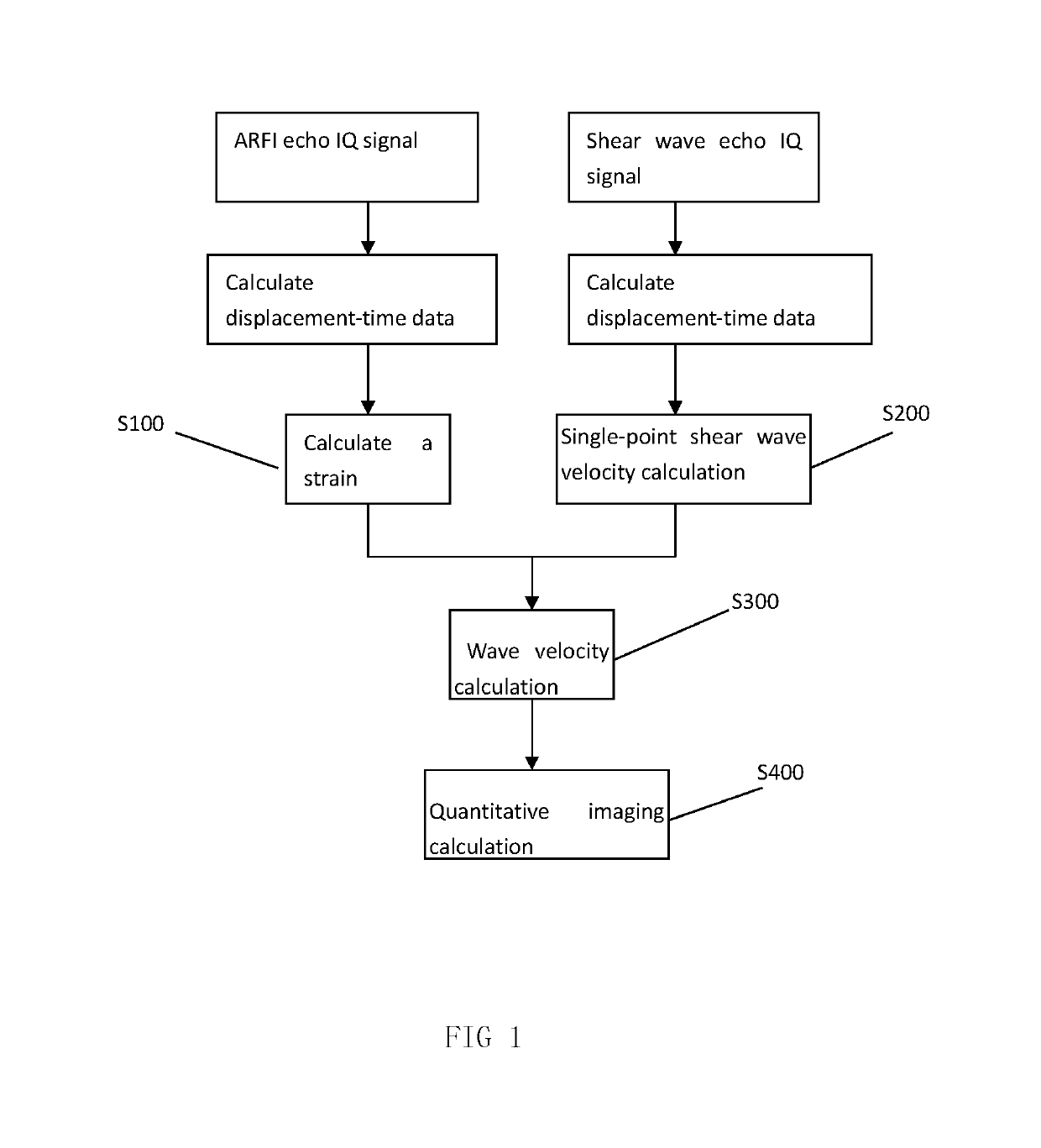
[0048]Embodiment 1: as shown in FIG. 1, the present invention provides a quantitative shear wave elasticity imaging method, including the following steps:

[0049]S100: Perform ARFI detection, and obtain a strain εref at a specified position in a focus region.

[0050]S200: Perform shear wave detection, and obtain a single-point shear wave velocity cref at the specified position in the focus region.

[0051]There is no special requirement for the execution sequence of steps S100 and S200. For example, S100 may be executed first, then S200 may be executed, or S200 may be executed first, and then S1...


PUM


Abstract

A quantitative shear wave elasticity imaging method and system relates to the technical field of medical ultrasound imaging. The provided ultrasound quantitative elasticity imaging method and system are based on a sliding window linear fitting strain and use a two-dimensional linear fitting shear wave velocity detection algorithm, and thus, the anti-noise capability is stronger, and the result is more reliable. Moreover, where the load of an ultrasonic front-end storage and transmission module is not additionally in-creased, global ultrasonic quantitative elasticity imaging is realized, thereby significantly reducing the design difficulty of the ultrasound quantitative elasticity imaging system and the device cost.

Description

CROSS-REFERENCE TO RELATED APPLICATIONS[0001]This application claims priority to and is a continuation of PCT / CN2017 / 107120, filed on Oct. 20, 2017, which claims priority to Chinese Patent Application Nos. 201610973108.8, filed on Nov. 4, 2016, and 201610973110.5, filed on November 4, 2016, the entireties of which are hereby incorporated by reference.TECHNICAL FIELD[0002]The present invention relates to the technical field of medical ultrasound imaging, and in particular, to a quantitative shear wave elasticity imaging method and system.BACKGROUND ART[0003]Clinically, changes in the mechanical characteristics of human tissues are often the most important early warning signals for tumor diseases. As the tumor grows, the viscoelasticity of the pathological tissue changes significantly compared with the normal tissue. For example, the breast cancer is approximately 90 times different from the surrounding normal tissue. The shear moduli of elasticity of fibrous tissues, non-invasive can...

Claims


Application Information

Patent Timeline
Patent Type & Authority Applications(United States)
IPC IPC(8): A61B8/00A61B8/08A61B8/14
CPCA61B8/54A61B8/485A61B8/14A61B8/085A61B8/08
Inventor YIN, HAOSHI, DANLIU, XIYAOLIU, DONGQUAN
Owner SASET CHENGDU TECH LTD
Why Eureka
Try Eureka