Method for measurement and control of intracular VEGF concentration

a technology of intracular veins and vegf, which is applied in the direction of instruments, material analysis, biological material analysis, etc., can solve the problems of short half-life of anti-vegf molecules in the human eye, uncontrolled formation of new blood vessels, and impede repeated injections to provide sustained vegf blockad

Inactive Publication Date: 2019-08-29
JUSTUS LIEBIG UNIV
View PDF1 Cites 2 Cited by
  • Summary
  • Abstract
  • Description
  • Claims
  • Application Information

AI Technical Summary

Problems solved by technology

Most retinal neovascular disorders are caused by upregulation of vascular endothelial growth factor (VEGF) expression, which leads to an uncontrolled formation of new, immature blood vessels in the eye.
The pivotal disadvantage of all anti-VEGF molecules is the short half-life in the human eye (e.g., t1 / 2=7.19 days for ranibizumab).
This impedes repeated injections to provide sustained VEGF blockade.
However, such repeated injections of anti-VEGF molecules are very expensive and may have severe side effects like intraocular inflammation or retinal detachment.
However, these methods have some major disadvantages: First, it is not clear whether the VEGF concentration in blood correlates with the VEGF concentration in the eye; second, dotting of the eye may cause severe afflictions like endophthalmitis that may lead to complete loss of sight; and third, all commercially available ELISA formats need high sample volumes (>5 μl) and have a detection limit in the range of picogram per milliliter (pg / ml).

Method used


Image

  • Method for measurement and control of intracular VEGF concentration
  • Method for measurement and control of intracular VEGF concentration
  • Method for measurement and control of intracular VEGF concentration

Examples


example 1

tion of Intraocular VEGF Concentration In Vivo

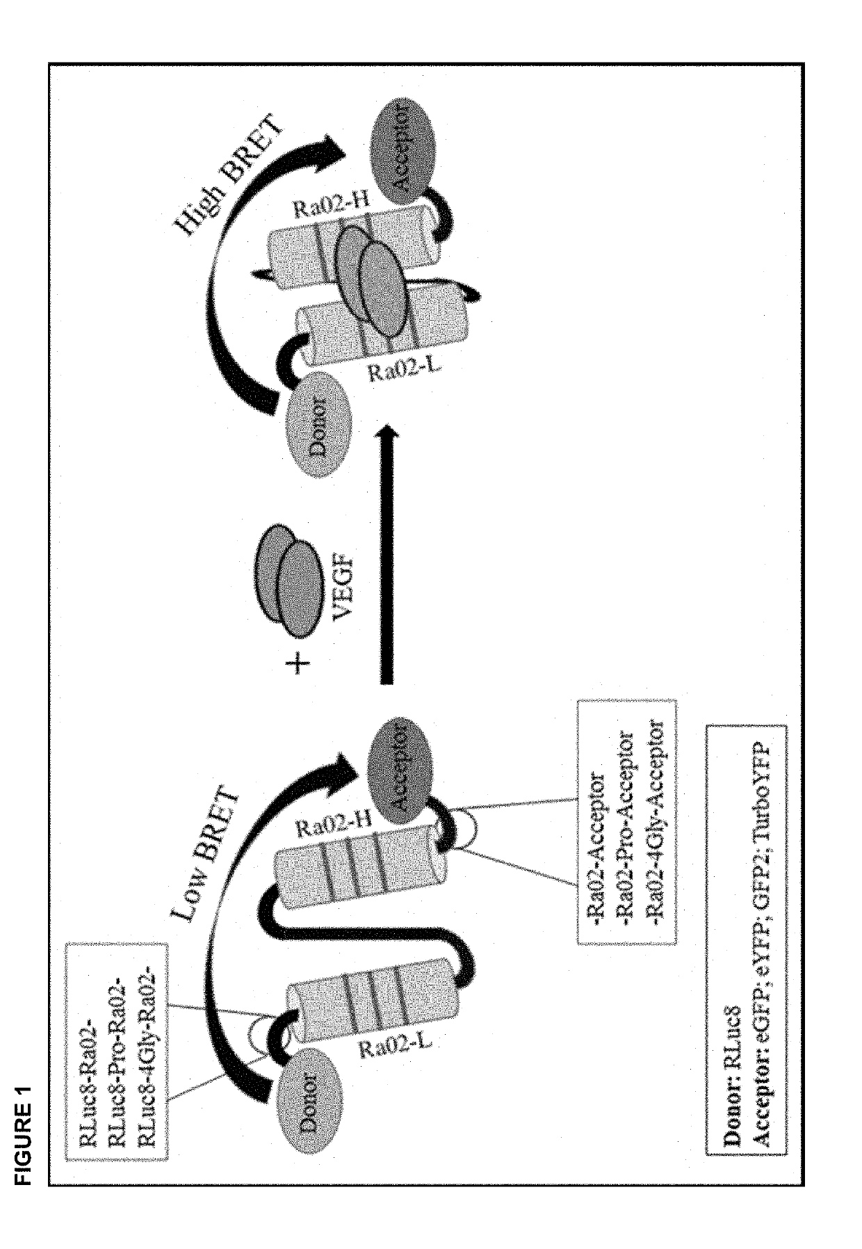
[0025]According to the present invention, the intraocular VEGF concentration in vivo is determined via the following steps. First, the substrate for Renilla luciferase, Coelenterazine, is administered to the patient whose VEGF concentration should be measured in the eye and to whom the VEGF-binding biosensor, for example RLuc8-Ra02-GFP2 (SEQ ID No.1), or RLuc8-Ra02-YFP, or RLuc8-Ra02-eYFP, or RLuc8-Ra02-TurboYFP, encapsulated in an insert, e.g. a microbead made from alginate, according to the present invention (FIG. 1, FIG. 2) has been implanted previously into the eye. Coelenterazine is commercially available, e.g. as EnduRen™ or ViviRen™ Live Cell Substrate (Promega), and is administered intravenously or orally, respectively, at doses that are known to the skilled worker. Subsequently, at an appropriate time after administration, the BRET ratio is measured using filters absorbing light at wavelengths of below 450 nm (signal from Renill...

example 2

of Anti-VEGF Synthesis In Vivo

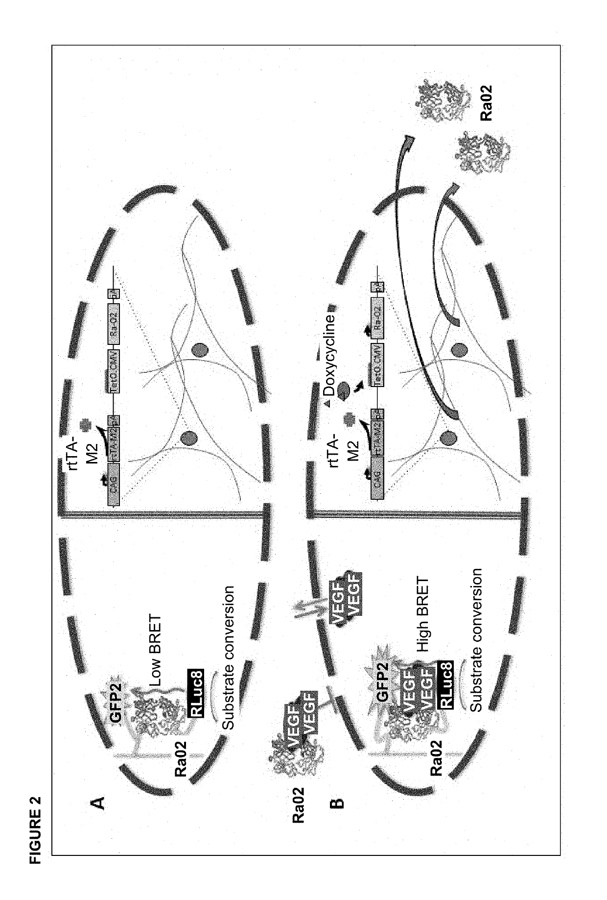
[0026]According to the invention described herein, the insert harbouring the VEGF-binding biosensor may additionally encapsulate a tetracycline-inducible (TetOn) vector encoding anti-VEGF molecules that is transduced into HEK-293 cells (FIG. 2). This vector is TetOn-Ra02 that has been previously described (Wimmer et al., Functional Characterization of AAV-Expressed Recombinant Anti-VEGF Single-Chain Variable Fragments In Vitro. J Ocul Pharmacol Ther. 2015 June; 31(5):269-76) (SEQ ID No. 2). Expression of anti-VEGF molecules from TetOn-Ra02 is induced by oral administration of doxycycline to the patient with doses according to the state of the art (0.5 to 10 mg / kg body weight). The newly synthesized anti-VEGF molecules bind to VEGF that is present in the eye and therefore reduces the concentration of free VEGF. The reduction of VEGF concentration can be determined at an appropriate time after administration of doxycycline by the method described in examp...

example 3

tion of Intraocular VEGF Concentration In Vitro

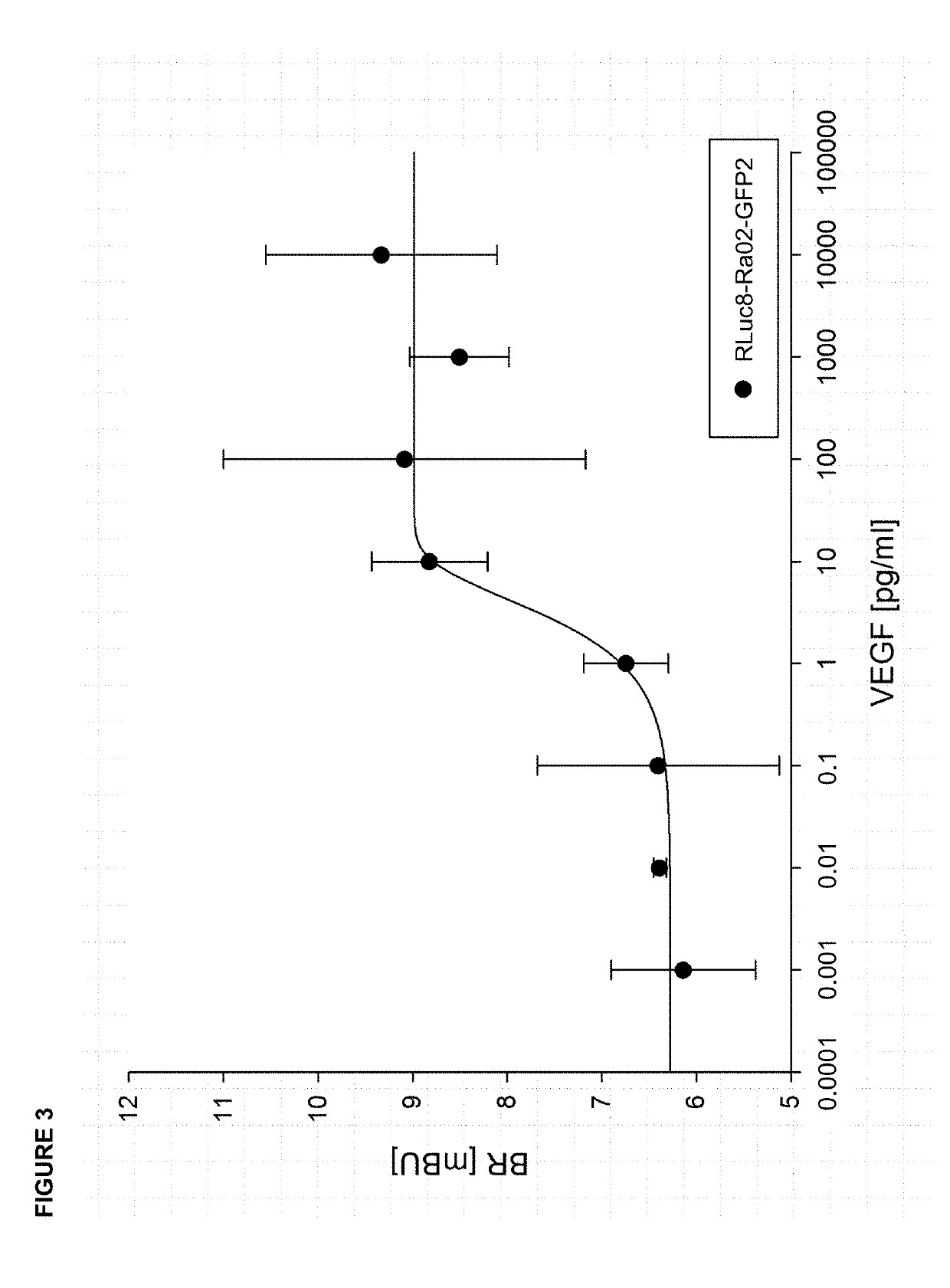
[0027]According to the invention presented herein, for determination of intraocular VEGF concentration in vitro, in a first step for example 6 μg of a vector encoding VEGF-binding biosensor molecules, for example RLuc8-Ra02-GFP2 (SEQ ID No. 1), are transfected in a 6-well with Lipofectamine 3000 (Invitrogen) into eukaryotic HEK-293 cells. This method is known to the skilled worker. RLuc8-Ra02-GFP2 comprises an anti-VEGF single chain variable fragment (anti-VEGF-scFv; Ra02) with Renilla luciferase (RLuc8) fused to its N-terminus and a fluorescent protein (GFP2) fused to its C-terminus. Afterwards, expression of RLuc8-Ra02-GFP2 biosensor molecules is allowed for 48 hours in an incubation chamber at 37° C. and 5% CO2. Expression is then verified by luciferase activity assay and fluorescence microscopy; both techniques are known from the state of the art. Subsequently, the newly synthesized biosensor molecules RLuc8-Ra02-GFP2 are isolated f...


PUM

PropertyMeasurementUnit
volumeaaaaaaaaaa
concentrationaaaaaaaaaa

Abstract

The invention describes a new method for in vivo measurement and control of intraocular VEGF concentration using bioluminescence resonance energy transfer (BRET) of a VEGF-binding biosensor. Furthermore, the method is suitable for highly sensitive in vitro determination of VEGF concentration from a small sample volume.

Description

BACKGROUND OF THE INVENTION[0001]Most retinal neovascular disorders are caused by upregulation of vascular endothelial growth factor (VEGF) expression, which leads to an uncontrolled formation of new, immature blood vessels in the eye. VEGF is a homodimeric heparin-binding glycoprotein with pro-angiogenic properties, stimulating migration and proliferation of micro- and macrovascular endothelial cells. The VEGF family consists of at least six subgroups named VEGF-A to VEGF-E and the placental growth factor. VEGF-A is the most prominent and potent isoform and key regulator in physiological angiogenesis as well as abnormal vascularization (neovascularization [NV]).[0002]The expression of VEGF is upregulated under hypoxic conditions and by various cytokines. Ocular diseases with NV as a typical pathological feature include the neovascular form of age-related macular degeneration (wet / exudative AMD), diabetic macular edema (DME) in patients with diabetic retinopathy, retinal vein occlus...

Claims


Application Information

Patent Timeline
Patent Type & Authority Applications(United States)
IPC IPC(8): A61K49/00G01N33/68
CPCA61K49/0047G01N2800/16G01N2333/475G01N33/6893G01N33/542A61K49/0021A61K49/00
Inventor LORENZ, BIRGITWIMMER, TOBIASSTIEGER, KNUT
Owner JUSTUS LIEBIG UNIV
Why Eureka
Try Eureka