
Human myosin peptides

a technology of myosin and peptides, which is applied in the field of human myosin peptides, can solve the problems that myocarditis is the leading cause of heart failure in people under 40, and achieve the effects of reducing autoantibodies to -myosin, reducing the proliferation of t cells, and improving left ventricular cardiac function

Inactive Publication Date: 2019-10-03
APITOPE INT
View PDF1 Cites 0 Cited by
  • Summary
  • Abstract
  • Description
  • Claims
  • Application Information

AI Technical Summary

Benefits of technology

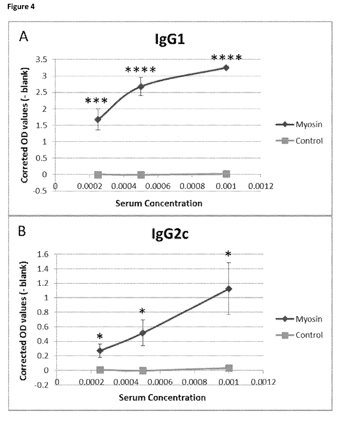
The present patent describes peptides from α-myosin that can reduce the production of autoantibodies to α-myosin, slow down the growth of T cells that target cardiac myosin, and improve heart function. These peptides can potentially prevent or treat autoimmune myocarditis.

Problems solved by technology

Myocarditis is the leading cause of heart failure in people under 40 years of age.

Method used


Image

  • Human myosin peptides
  • Human myosin peptides
  • Human myosin peptides

Examples


example 1

Materials and Methods

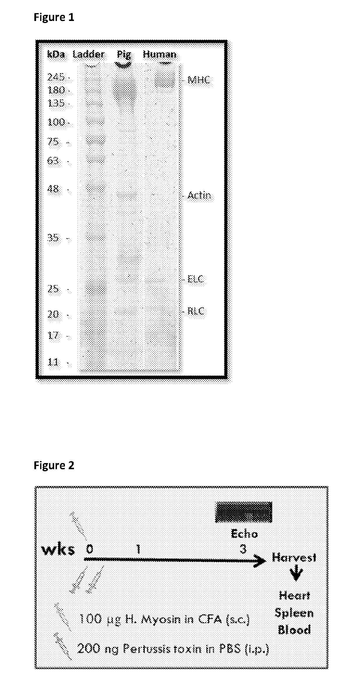
[0113]Myosin Extraction from Human Heart

[0114]A suitably sized chunk of the left ventricular wall was taken out, thawed and minced. Each gram of minced heart was homogenized in 10 ml of Solution A (0.4 M KCl; 0.15 M K2HPO4; 0.01 M Na4P2O7; 0.001 M MgCl2; 0.002 M DTT; pH:6.8 adjusted by KH2PO4; all chemicals used in this protocol were obtained from Sigma Aldrich) and centrifuged at 150,000×g for 1 hour to clear the muscle residues and cellular debris. The supernatant was diluted with >20 volumes of 2 mM DTT to precipitate filamentous myosin which was pelleted by subsequent centrifugation at 50,000×g for 20 mins. The pellet was re-suspended in 10 ml of Solution B (0.3 M KCl; 0.004 M MgCl2; 0.025 M imidazole; 0.01 M DTT; 0.001 M EGTA; pH:7.4 adjusted by HCl) and centrifuged at 43,000×g for 30 mins to remove actin. The supernatant was diluted with >7 volumes of 2 mM DTT and centrifuged at 12,000×g for 20 minutes. The pellet was re-suspended in 6 ml 0.3 M KCl, 0.01 M...


PUM

PropertyMeasurementUnit
Fractionaaaaaaaaaa
Fractionaaaaaaaaaa
Pharmaceutically acceptableaaaaaaaaaa

Abstract

The present invention provides a peptide at least partially derivable from human α-myosin which peptide and the use of such peptides for the prevention or suppression of autoantibody formation in myocarditis.

Description

FIELD OF THE INVENTIONBackground to the Invention[0001]Myocarditis is the leading cause of heart failure in people under 40 years of age. Up to one fifth of those affected by an acute episode will progress towards chronic disease leading to dilated cardiomyopathy (DCM). Acute myocarditis can result from infection by viruses including coxsackevirus B3 (CVB3); evidence of prior infection is found in approximately 50% of patients with DCM. Viruses such as CVB3 have a direct cytopathic effect on cardiac tissue. Strong evidence suggests, however, that individuals who progress towards chronic myocarditis do so because they develop an autoimmune response to antigens of cardiac muscle.[0002]Circulating antibodies to cardiac tissue antigens, including myosin, are associated with chronic myocarditis and the severity of cardiac disease is directly related to levels of autoantibodies in the affected tissue. Organ specific IgG antibodies were found in biopsies from patients with DCM. B cells sec...

Claims


Application Information

Patent Timeline
IPC IPC(8): C07K7/08A61P9/00A01K67/027A61K49/00
CPCA01K2227/105A01K2207/10A01K67/0278A61P9/00A61K49/0008A01K2267/0325C07K7/08A61K38/00A61K39/00C07K14/4716A61K39/0008A61K2039/577A61P29/00
Inventor SELLI, MEHMETWRAITH, DAVID
Owner APITOPE INT
Learn More