
Optical Detection And Measurement Of Hematocrit And Free Hemoglobin Concentration

a technology of free hemoglobin and optical detection, which is applied in the field of optical detection and measurement of free hemoglobin concentration, can solve the problems of erroneous measurement, low light transmission through fluid containing even low quantities of red blood cells, and easy disturbance of the fluid,

Active Publication Date: 2019-10-17
FENWAL
View PDF0 Cites 3 Cited by
  • Summary
  • Abstract
  • Description
  • Claims
  • Application Information

AI Technical Summary

Benefits of technology

The present subject matter describes a method and device for measuring the hematocrit or free hemoglobin concentration of a fluid. The device includes a broadband light source that emits light and a vessel containing the fluid. The light is reflected by the fluid and received by an optical spectrometer. The optical spectrometer analyzes the received light and determines the main wavelength of the light, which is correlated to the hematocrit or free hemoglobin concentration of the fluid. An output is generated that indicates the hematocrit or free hemoglobin concentration of the fluid.

Problems solved by technology

However, light transmission through fluid containing even low quantities of red blood cells (i.e., fluids having a relatively low hematocrit) may be minimal due to significant scattering of the light.
Another possible disadvantage of such transmission-based approaches is that they require access to opposing sides of a fluid path, which may not be available for certain fluid paths in certain systems.
While such techniques are capable of producing reliable results, they may be susceptible to disturbances, such as when air bubbles are present in the fluid.
When light being transmitted through a fluid encounters air bubbles, the light is scattered, which reduces the amount of light transmitted through the fluid to a light detector and may lead to erroneous measurements.
Additionally, plasma or supernatant obtained from a product after separation or during storage is conventionally evaluated using specific free hemoglobin measurement assays, which may be time-consuming and costly.

Method used


Image

  • Optical Detection And Measurement Of Hematocrit And Free Hemoglobin Concentration
  • Optical Detection And Measurement Of Hematocrit And Free Hemoglobin Concentration
  • Optical Detection And Measurement Of Hematocrit And Free Hemoglobin Concentration

Examples


Embodiment Construction

[0032]The embodiments disclosed herein are for the purpose of providing a description of the present subject matter, and it is understood that the subject matter may be embodied in various other forms and combinations not shown in detail. Therefore, specific designs and features disclosed herein are not to be interpreted as limiting the subject matter as defined in the accompanying claims.

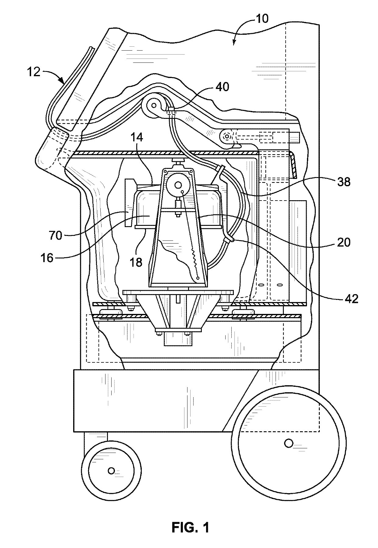
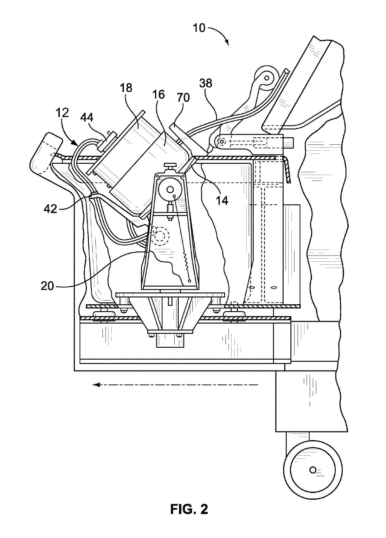
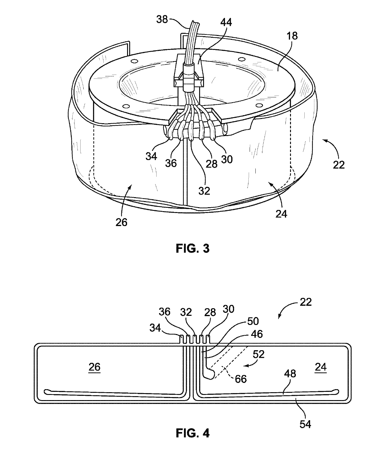
[0033]FIGS. 1 and 2 show a biological fluid processing system that may be used in practicing the hematocrit and free hemoglobin concentration measurement principles of the present disclosure. The system is currently marketed as the AMICUS® separator of Fenwal, Inc. and is described in greater detail in U.S. Pat. No. 5,868,696, which is hereby incorporated herein by reference. The system can be used for processing various fluids, but is particularly well suited for processing whole blood, blood components, or other suspensions of biological cellular materials. While hematocrit and free hemoglobin co...


PUM

PropertyMeasurementUnit
angleaaaaaaaaaa
angleaaaaaaaaaa
angleaaaaaaaaaa

Abstract

A colorimetric optical sensor device includes a broadband light source configured to emit a light that is exposed to a fluid in a vessel. At least a portion of the light is reflected off of the fluid and received by an optical spectrometer. The optical spectrometer analyzes at least a portion of the received light to determine a main wavelength of the light. A controller correlates the main wavelength to a corresponding hematocrit or free hemoglobin concentration and generates an output indicative of the hematocrit or the free hemoglobin concentration of the fluid. The hematocrit or free hemoglobin concentration information may be used by a separation assembly in which the colorimetric optical sensor device may be incorporated to modify a separation procedure in which the monitored fluid is to be separated or is a component or constituent of a previously separated biological fluid.

Description

CROSS-REFERENCE TO RELATED APPLICATIONS[0001]This application claims the benefit of and priority of U.S. Provisional Patent Application Ser. No. 62 / 657,397, filed Apr. 13, 2018, the contents of which are incorporated by reference herein.BACKGROUNDField of the Disclosure[0002]The present disclosure relates to optical detection and measurement of hematocrit and free hemoglobin concentration. More particularly, the present disclosure relates to non-invasive colorimetric approaches to measuring hematocrit and free hemoglobin concentration using diffuse reflectance spectroscopy.Description of Related Art[0003]Various blood processing systems now make it possible to collect particular blood constituents, instead of whole blood, from a blood source such as, but not limited to, a container of previously collected blood or other living or non-living source. Typically, in such systems, whole blood is drawn from a blood source, the particular blood component or constituent is separated, remove...

Claims


Application Information

Patent Timeline
Patent Type & Authority Applications(United States)
IPC IPC(8): A61B5/145A61B5/1455G01N33/49G01N21/25G01N33/487A61M1/36
CPCG01N21/251A61M2202/0415A61M2202/0433A61B5/14535A61B5/1455G01N33/48778G01N2201/08A61M2205/3306G01N2201/062A61M1/3693G01N33/491G01N21/255G01N21/31G01N21/49A61B5/15003A61B5/150992A61M1/362265A61M1/36224A61M1/362261
Inventor KUSTERS, BENJAMIN E.MIN, KYUNGYOON
Owner FENWAL
Learn More