
Optical sectioning apparatus using advanced mirau optical interference microscopy
a technology of optical interference and optical sectioning, which is applied in the field of optical sectioning apparatus, can solve the problems of damage to the tissue structure, time-consuming pathologist's examination of frozen sections, and inability to guarantee the removal of tumors
- Summary
- Abstract
- Description
- Claims
- Application Information
AI Technical Summary
Benefits of technology
Problems solved by technology
Method used
Image

Examples
Embodiment Construction
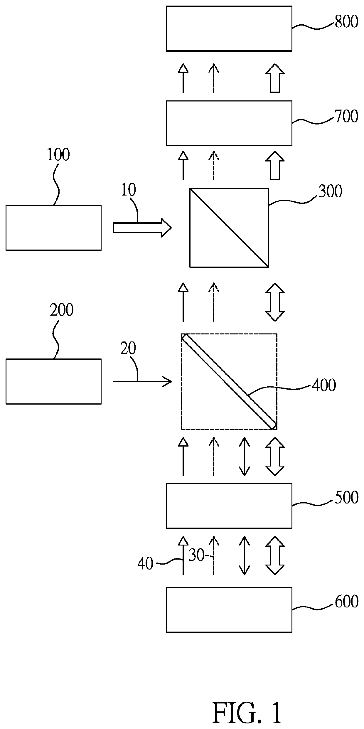
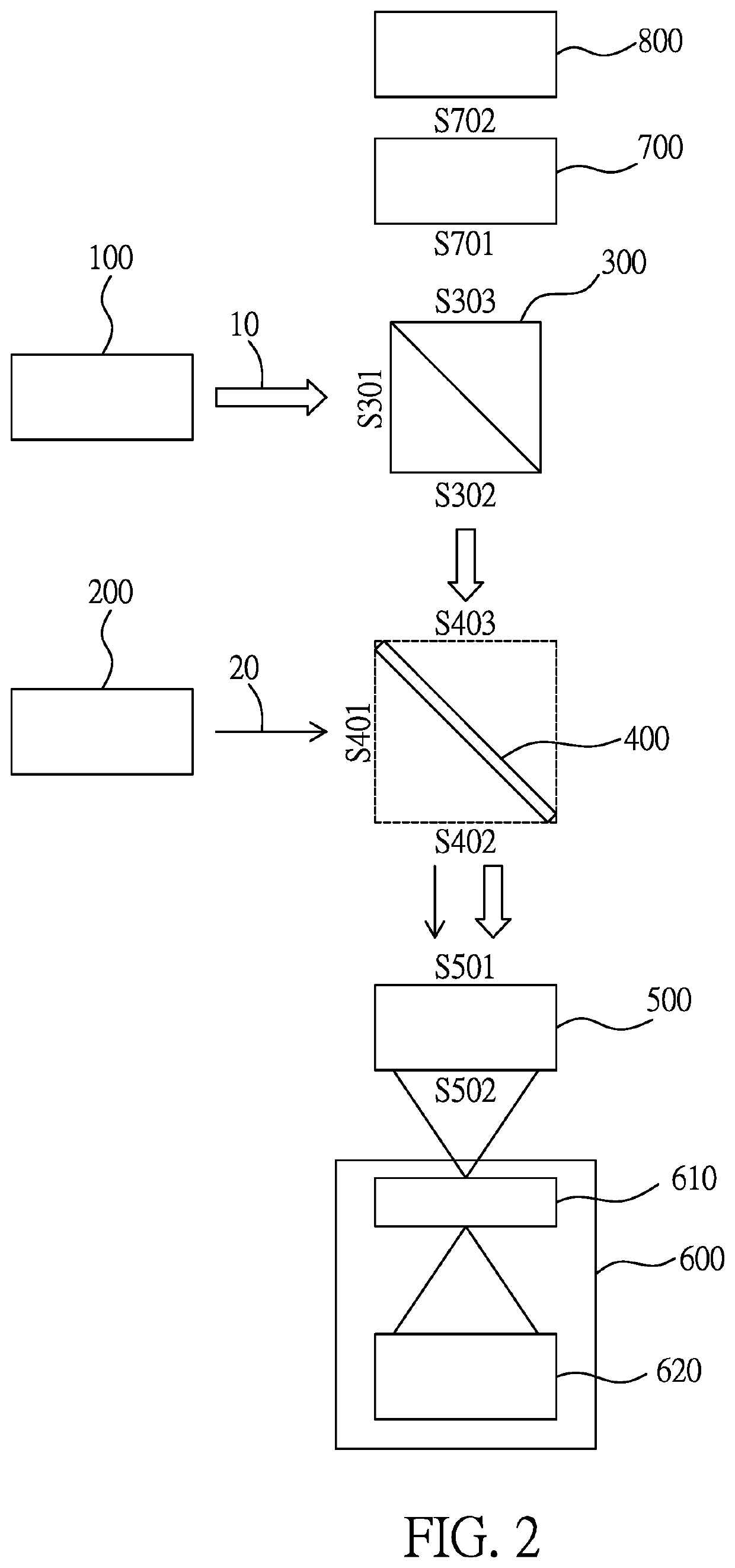
[0037]Please refer to FIG. 1-2, in which, FIG. 1 illustrates a block diagram of an optical sectioning apparatus combining Mirau optical interference microscopy and fluorescence microscopy according to one embodiment of the present invention, and FIG. 2 illustrates a beam splitting operation and a focusing operation of the optical sectioning apparatus of FIG. 1.
[0038]As illustrated in the figures, the optical sectioning apparatus combining Mirau optical interference microscopy and fluorescence microscopy includes: a wide band light source apparatus 100, a short wavelength light source apparatus 200, an optical circulator 300, a first dichroic splitter 400, a Mirau optical interference objective lens 500, a sample carrier unit 600, a projection lens 700, and a sensor unit 800.
[0039]The wide band light source apparatus 100 is used to generate a wide band light beam 10 (indicated by a hollow arrow); the short wavelength light source apparatus 200 is used to generate a short wavelength l...
PUM
 Abstract
Description
Claims
Application Information