
Cancer classifier models, machine learning systems and methods of use

Pending Publication Date: 2020-01-02
20 20 GENESYSTEMS INC
View PDF0 Cites 18 Cited by
  • Summary
  • Abstract
  • Description
  • Claims
  • Application Information

AI Technical Summary

Benefits of technology

The patent text describes a computer-implemented system for predicting an increased risk of cancer in a patient. The system uses a machine learning system to analyze biomarker data from the patient, such as age and gender, and assign the patient to a risk category based on the levels of biomarkers measured in the sample. The system can also predict which organ system the cancer is likely to develop in the patient. The system can be iteratively improved through the use of new training data. The technical effect of this patent is the development of a more accurate and reliable method for predicting cancer risk in patients, which can help healthcare professionals provide earlier and more targeted diagnostic testing for cancer.

Problems solved by technology

Unfortunately, the costs of conducting large prospective studies for screening tools is outweighed by reasonably anticipated financial returns so these large prospective studies are almost never done by the private sector and are only occasionally sponsored by governments.
As a result, the use paradigms for blood testing for the early detection of most cancers has progressed little in several decades.
In the United States, for example, PSA remains the only widely utilized blood test for cancer screening and even its utilization has become controversial.
In other parts of the world, especially the Far East, blood tests for detecting various cancers is more commonplace but there is little standardization or empirical methods to ascertain or improve the accuracy of such testing in those parts of the world.
Cancer detection poses significant technical challenges as compared to detecting viral or bacterial infections since cancer cells, unlike viruses and bacteria, are biologically similar to and hard to distinguish from normal, healthy cells.
For this reason, tests used for the early detection of cancer often suffer from higher numbers of false positives and false negatives than comparable tests for viral or bacterial infections or for tests that measure genetic, enzymatic, or hormonal abnormalities.
This often causes confusion among healthcare practitioners and their patients leading in some cases to unnecessary, expensive, and invasive follow-up testing while in other cases to a complete disregard for follow-up testing resulting in cancers being detected too late for useful intervention.
However, unless the sensitivity and specificity of diagnosis approaches 99%, a level not obtainable for most cancer tests, such binary outputs can be highly misleading or inaccurate.
Detecting early stage cancer is also challenging due to factors associated with the modern-day practice of medicine.
Primary care providers in particular, see a high volume of patients per day and the demands of healthcare cost containment has dramatically shortened the amount of time they can spend with each patient.
Accordingly, physicians often lack sufficient time to take in depth family and lifestyle histories, to counsel patients on healthy lifestyles, or to follow-up with patients who have been recommended testing beyond that which is provided in their office practice.
Although decision-making systems have been developed, such systems are not widely used in medical practice because these systems suffer from limitations that prevent them from being integrated into the day-to-day operations of health organizations.
For example, decision-making systems may provide an unmanageable volume of data, rely on analysis that is marginally significant, and not correlate well with complex multimorbidity (Greenhalgh, T. Evidence based medicine: a movement in crisis?
Also, the systems are difficult to interact with (Berner, 2006; Shortliffe, 2006).
The entry of patient data is difficult, the list of diagnostic suggestions may be too long, and the reasoning behind diagnostic suggestions is not always transparent.
Further, the systems are not focused enough on next actions, and do not help the clinician figure out what to do to help the patient (Shortliffe, 2006).

Method used


Image

  • Cancer classifier models, machine learning systems and methods of use
  • Cancer classifier models, machine learning systems and methods of use
  • Cancer classifier models, machine learning systems and methods of use

Examples


example 1a

t of a Multi-Marker Model for Classifying Asymptomatic Patients as to Developing Cancer: “Pan Cancer” Test

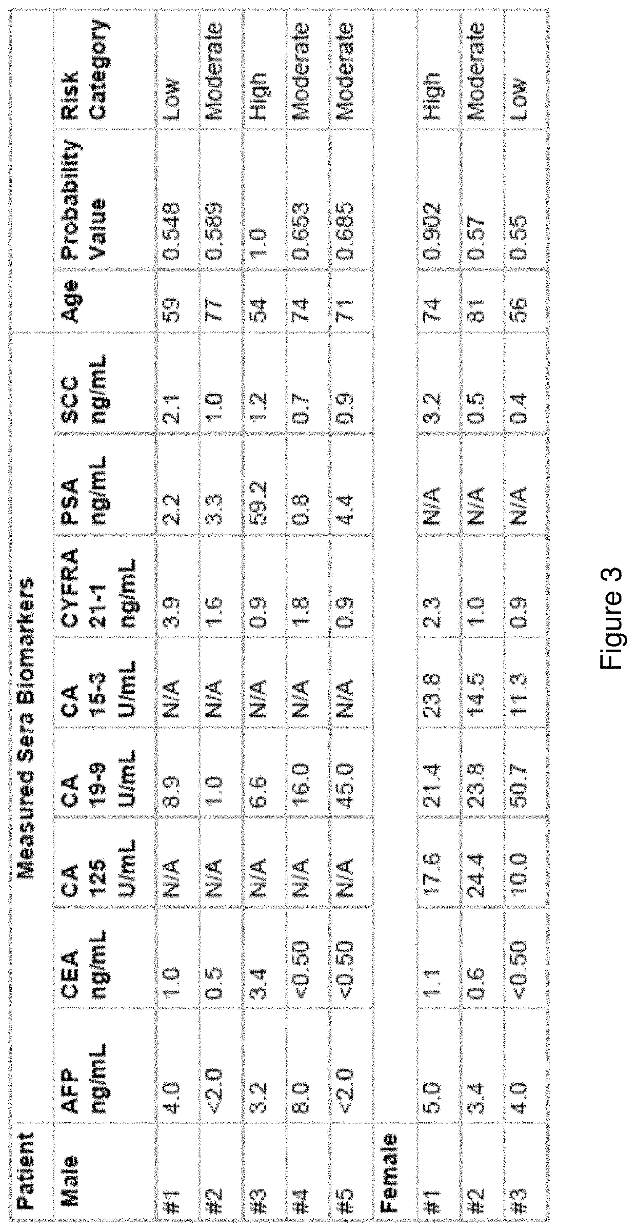
[0159]Provided herein is a multi-marker classification model and method for identifying asymptomatic patients with an increased risk for developing cancer. That risk can be categorized as “low”, “medium / moderate” or “high risk” for developing cancer, wherein the ranges for those categories may be based on, for example, probability of developing cancer within 6 months to a year, wherein the probability is measured against baseline level of cancer in the heterogenous population. It is understood in the art, that the rate of cancer is about 1% in the general population. The prevalence of cancer in the cohort used to develop the present Pan Cancer test was about 1.5%. See the below examples for more detail on the use of the test and probability values. The development of the classifier model, and the selection of markers (both blood and clinical parameters) may be based on a combina...

example 1b

t of a Multi-Marker Model for Classifying Asymptomatic Patients as to Developing Cancer: Inclusion of Clinical Factor “Age” in Model

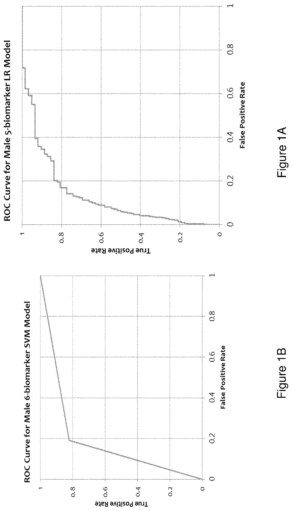
[0180]Disclosed herein is an improved multi-marker model for classifying asymptomatic patients as to having or developing cancer. The above classifier model using only a panel of measured biomarkers was previously published wherein the performance of a Receiver Operating Characteristic (ROC) curve for the cohort of males was very low; sensitivity value of 0.515 and a specificity value of 0.851. The cohort of females had an even lower performance of a ROC curve with a sensitivity value of 0.345 and a specificity value of 0.880. See Tables 7 and 8 of Wang H. Y., Hsieh C. H., Wen C. N., Wen Y. H., Chen C. H. and Lu J. J., “Cancers Screening in an Asymptomatic Population by Using Multiple Tumour Markers” PLoS One, Jun. 29, 2016. In other words, the previous classifier model using only measured sera biomarkers was acceptable for excluding the risk of cancer ...

example 2

nt of a Model for Predicting Organ System-Based Malignancy for Individuals in the “High Risk” and “Moderate Risk” Category Based on the Pan Cancer Test

[0182]Provided herein are techniques for predicting organ system-based malignancy for a patient with an increased risk of having cancer as identified in Example 1. That information can then be used to refer patients to a specialist for more invasive diagnostic testing.

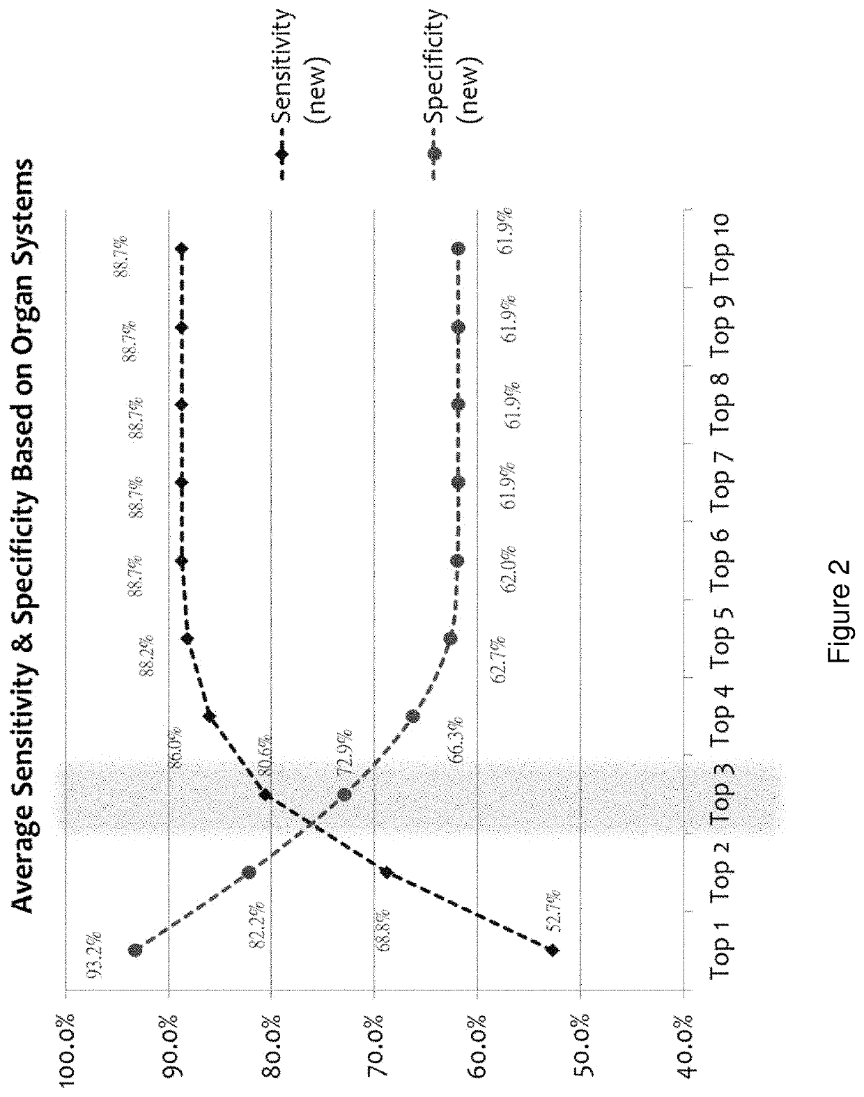
[0183]Using the entire cohort of cancer subjects (n=186) and the same six (or 5 for female individuals) biomarker measurements along with age and gender, we applied a model comprising a pattern recognition algorithm, and a k-Nearest Neighbors algorithm (kNN) employing a leave-one-out evaluation method to predict the top 1, 2, 3, 4, 5, 6, 7, 8, 9, or 10 cancers for each sample. The accuracies are reported in Table 5 and reflect the percentage of cases of each cancer type that were found in the top N (N=10 for Table 5) predicted cancers. Clearly, the accuracy of prediction...


PUM


Abstract

Disclosed herein are classifier models, computer implemented systems, machine learning systems and methods thereof for classifying asymptomatic patients into a risk category for having or developing cancer and / or classifying a patient with an increased risk of having or developing cancer into an organ system-based malignancy class membership and / or into a specific cancer class membership.

Description

CROSS-REFERENCE TO RELATED APPLICATIONS[0001]This application claims the benefit of U.S. Provisional Patent Application No. 62 / 692,683, filed on 30 Jun. 2018, the content of which is incorporated herein by reference in its entirety.FIELD OF THE INVENTION[0002]This application pertains generally to classifier models generated by a machine learning system, trained with longitudinal data, for identifying asymptomatic patients with an increased risk for developing cancer and the type of cancer, especially in an otherwise asymptomatic or vaguely symptomatic patient.BACKGROUND OF THE INVENTION[0003]For many types of cancers, patient outcomes improve significantly if surgery and other therapeutic interventions commence before the tumor has metastasized. Accordingly, imaging and diagnostic tests have been introduced into medical practice in an attempt to help physicians detect cancer early. These include various imaging modalities such as mammography as well as diagnostic tests to identify ...

Claims


Application Information

Patent Timeline
IPC IPC(8): G16B40/20G06N20/00G16H50/20G16H50/30
CPCG16B40/20G16H50/20G16H50/30G06N20/00G16H50/70G16B20/00G06N20/20G06N20/10G06N5/01G06N3/045
Inventor COHEN, JONATHANDOSEEVA, VICTORIASHI, PEICHANG
Owner 20 20 GENESYSTEMS INC
Learn More